Review
. 2010 Apr;9(4):425-37.
doi: 10.1016/S1474-4422(10)70040-5.

Progressive multifocal leukoencephalopathy and other disorders caused by JC virus: clinical features and pathogenesis

Affiliations
Review

Progressive multifocal leukoencephalopathy and other disorders caused by JC virus: clinical features and pathogenesis

Chen S Tan et al. Lancet Neurol. 2010 Apr.

Abstract

Progressive multifocal leukoencephalopathy (PML) is a rare but often fatal brain disease caused by reactivation of the polyomavirus JC. Knowledge of the characteristics of PML has substantially expanded since the introduction of combination antiretroviral therapy during the HIV epidemic and the development of immune reconstitution inflammatory syndrome (IRIS) in patients with PML. Recently, the monoclonal antibodies natalizumab, efalizumab, and rituximab--used for the treatment of multiple sclerosis, psoriasis, haematological malignancies, Crohn's disease, and rheumatic diseases--have been associated with PML. Additionally, the JC virus can also lead to novel neurological disorders such as JC virus granule cell neuronopathy and JC virus encephalopathy, and might also cause meningitis. The increasingly diverse populations at risk and the recent discovery of the presence of the JC virus in the grey matter invite us to reappraise the pathogenesis of this virus in the CNS.


Figures

Figure 1
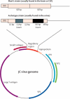
Figure 1. JC virus Genome
JCV genome is composed of a 5kb of double stranded circular DNA. The coding region includes the small t, large T antigens, the capsid proteins VP1, VP2, and VP3, and the agnoprotein (Agno). The non-coding regulatory region (RR) detected in the brain or CSF of PML patients usually consists of tandem repeats of a 98 bp element, as in the Mad-1 strain, and most isolates from urine of healthy and PML patients alike, are similar to the archetype RR. JCV archetype has one 98 bp element with a 23 bp insert (gray box) and a 66 bp insert (black box).
Figure 2
Figure 2. Cerebellar lesions in a patient with classic PML
A 69 years old man with CLL, treated successively with combination chemotherapy and rituximab presented with 6 weeks history of progressive decline in right hand and leg coordination, and new onset of severe vertigo and emesis. MRI showed an area of hyperintense signal in the right cerebellar hemisphere and cerebellar peduncle on FLAIR (A, arrow), which appeared hypointense and was devoid of contrast enhancement on T1-weighted image (B, arrow). JCV PCR was positive in CSF. Rituximab was discontinued and he was started on mirtazapine 15mg per day. His neurological deficit continued to progress, including dysphagia and repeat MRI performed 10 days later showed extension of the lesions to the brain stem and the left cerebellar hemisphere on FLAIR image (C, arrow) and absence of enhancement on T1-weighted image (D, arrow). He passed away 3 months after PML diagnosis.
Figure 3
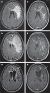
Figure 3. PML-IRIS in an HIV+ patient
A 40 yo man with HIV infection, who presented with progressive onset of word finding difficulties and right hemiparesis followed by seizure, 4 days after starting cART. PCR was positive for JCV in the CSF peripheral CD4 count was 468 cells/ul. MRI performed at another hospital reported a 3 cm focus of abnormal increased signal on FLAIR sequences in the left frontal subcortical white matter, surrounded by linear and punctate foci of enhancement at the margins of the lesion. This lesion extended into the left corona radiata, the corpus callosum and the right frontal white matter. MRI performed at our hospital 3 week after the initial one showed lesions in FLAIR (A, arrows) and contrast enhancement in T1-weighted image post gadolinium injection (B, arrowheads). His aphasia improved progressively with addition of ritonavir to his cART regimen. His CD4 count increased to 558 cells/ul and his HIV plasma viral load was undetectable. He then presented with worsening aphasia. MRI performed 2 and a half month after onset of initial symptoms showed enlargement of the lesions in the left hemispheric white matter and the corpus callosum in FLAIR (C, arrows) which displayed intense contrast enhancement in T1-weighted images (D, arrowheads) as well as mass effect, right to left shift and subfalcine herniation. He was treated with dexamethasone 6 mg three times a day, tapered over 2 weeks, and cART was discontinued for two weeks. All neurological symptoms progressively improved and 2 and a half year later, he has no residual weakness and only minor word finding difficulties. MRI showed leukomalacia and atrophy of the left frontal lobe with dilatation of the left lateral ventricule in FLAIR (E, arrows) and absence of contrast enhancement in T1-weighted image (F, arrowheads). His CD4 count was 669/ul and HIV plasma viral load continue to be undetectable.
Figure 4
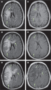
Figure 4. A natalizumab-treated MS patient with worsening PML after plasma exchange
A 35 years old woman with seven years history of relapsing remitting MS was treated with natalizumab monotherapy after failure with other treatments. After almost two years of monthly infusions, she presented with dysarthria, left-sided numbness and weakness. MRI of the brain showed a large right frontal lobe white matter hyperintense lesion on FLAIR, extending through the corpus callosum (A, arrows) which did not enhance on T1-weighted images (B, arrows). The patient was treated with plasma exchange every other day for 5 days. Nevertheless, symptoms worsened and repeat MRI 3 weeks later showed progression of the lesions on FLAIR with slight mass effect on the right lateral ventricle (C) without evidence of enhancement on T1-weighted images (D). The patient’s neurological condition worsened and follow up MRI done 3 ½ weeks later showed extension of the lesions in the right frontal and parietal lobes, as well as in the left frontal lobe, associated with edema and further mass effect on the lateral ventricles in FLAIR (E) which displayed faint areas of linear enhancement on T1 weighted images (F).
Figure 5
Figure 5. Histological spectrum of JCV infection of the central nervous system
A) Classic PML: demyelinating lesion of the white matter (arrow) surrounded by multiple JCV-infected glial cells (arrowheads). B) JCV GCN: JCV infection of granule cell neurons (arrows). C) PML-IRIS: marked lymphocytes penetrating the perivascular region (arrows). D) JCV encephalopathy: JCV infected (arrow) hemispheric cortical neurons (arrowhead). Panels A, B, and D courtesy of Dr. Christian Wurthrich. Pannel C courtesy of Dr Francoise Gray)

Comment in

Similar articles

Cited by

References

    1. Koralnik IJ. Progressive multifocal leukoencephalopathy revisited: Has the disease outgrown its name? Ann Neurol. 2006 Aug;60(2):162–73. - PubMed
    1. Eng PM, Turnbull BR, Cook SF, Davidson JE, Kurth T, Seeger JD. Characteristics and antecedents of progressive multifocal leukoencephalopathy in an insured population. Neurology. 2006 Sep 12;67(5):884–6. - PubMed
    1. Holman RC, Janssen RS, Buehler JW, Zelasky MT, Hooper WC. Epidemiology of progressive multifocal leukoencephalopathy in the United States: analysis of national mortality and AIDS surveillance data [see comments] Neurology. 1991;41(11):1733–6. - PubMed
    1. Koralnik IJ, Schellingerhout D, Frosch MP. Case records of the Massachusetts General Hospital. Weekly clinicopathological exercises. Case 14-2004. A 66-year-old man with progressive neurologic deficits. N Engl J Med. 2004 Apr 29;350(18):1882–93. - PubMed
    1. Molloy ES, Calabrese LH. Progressive multifocal leukoencephalopathy: a national estimate of frequency in systemic lupus erythematosus and other rheumatic diseases. Arthritis Rheum. 2009 Dec;60(12):3761–5. - PubMed

Publication types

MeSH terms