The indeterminate adrenal lesion
- PMID: 20299300
- PMCID: PMC2842175
- DOI: 10.1102/1470-7330.2010.0012
The indeterminate adrenal lesion
Abstract
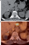
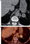
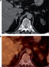
With the increasing use of abdominal cross-sectional imaging, incidental adrenal masses are being detected more often. The important clinical question is whether these lesions are benign adenomas or malignant primary or secondary masses. Benign adrenal masses such as lipid-rich adenomas, myelolipomas, adrenal cysts and adrenal haemorrhage have pathognomonic cross-sectional imaging appearances. However, there remains a significant overlap between imaging features of some lipid-poor adenomas and malignant lesions. The nature of incidentally detected adrenal masses can be determined with a high degree of accuracy using computed tomography (CT) and magnetic resonance imaging (MRI) alone. Positron emission tomography (PET) is also increasingly used in clinical practice in characterizing incidentally detected lesions. We review the performance of the established and new techniques in CT, MRI and PET that can be used to distinguish benign adenomas and malignant lesions of the adrenal gland.
Figures




Similar articles
-
Imaging evaluation of the non-functioning indeterminate adrenal mass.Trends Endocrinol Metab. 2004 Aug;15(6):271-6. doi: 10.1016/j.tem.2004.06.012. Trends Endocrinol Metab. 2004. PMID: 15358280 Review.
-
The indeterminate adrenal mass in patients with cancer.Cancer Imaging. 2007 Oct 1;7 Spec No A(Special issue A):S100-9. doi: 10.1102/1470-7330.2007.9017. Cancer Imaging. 2007. PMID: 17921094 Free PMC article. Review.
-
Radiologic evaluation of incidentally discovered adrenal masses.Am Fam Physician. 2010 Jun 1;81(11):1361-6. Am Fam Physician. 2010. PMID: 20521756 Review.
-
Non-invasive evaluation of the incidentally detected indeterminate adrenal mass.Best Pract Res Clin Endocrinol Metab. 2005 Jun;19(2):277-92. doi: 10.1016/j.beem.2004.09.002. Best Pract Res Clin Endocrinol Metab. 2005. PMID: 15763701 Review.
-
Characterization of adrenal adenomas and metastases: correlation between unenhanced computed tomography and chemical shift magnetic resonance imaging.Acta Radiol. 2006 Feb;47(1):71-6. doi: 10.1080/02841850500405193. Acta Radiol. 2006. PMID: 16498936 Clinical Trial.
Cited by
-
Multiparametric PET/CT in oncology.Cancer Imaging. 2012 Sep 28;12(2):336-44. doi: 10.1102/1470-7330.2012.9007. Cancer Imaging. 2012. PMID: 23023069 Free PMC article. Review.
-
Prediction of 2-[18F]FDG PET-CT SUVmax for Adrenal Mass Characterization: A CT Radiomics Feasibility Study.Cancers (Basel). 2023 Jun 30;15(13):3439. doi: 10.3390/cancers15133439. Cancers (Basel). 2023. PMID: 37444549 Free PMC article.
-
Radiomics approach based on biphasic CT images well differentiate "early stage" of adrenal metastases from lipid-poor adenomas: A STARD compliant article.Medicine (Baltimore). 2022 Sep 23;101(38):e30856. doi: 10.1097/MD.0000000000030856. Medicine (Baltimore). 2022. PMID: 36197274 Free PMC article.
-
Artificial intelligence and radiomics applications in adrenal lesions: a systematic review.Ther Adv Urol. 2025 Aug 2;17:17562872251352553. doi: 10.1177/17562872251352553. eCollection 2025 Jan-Dec. Ther Adv Urol. 2025. PMID: 40761430 Free PMC article. Review.
-
Distinguishing between metastatic and benign adrenal masses in patients with extra-adrenal malignancies.Front Endocrinol (Lausanne). 2022 Sep 23;13:978730. doi: 10.3389/fendo.2022.978730. eCollection 2022. Front Endocrinol (Lausanne). 2022. PMID: 36246921 Free PMC article.
References
-
- Glazer HS, Weyman PJ, Sagel SS, Levitt RG, McClennan BL. Non-functioning adrenal masses: incidental discovery on computed tomography. AJR. 1982;139:81–5. - PubMed
-
- Bovio S, Cataldi A, Reimondo G, et al. Prevalence of adrenal incidentaloma in a contemporary computerized tomography series. J Endocrinol Invest. 2006;29:298–302. - PubMed
-
- Song JH, Chaudhry FS, Mayo-Smith WW. The incidental adrenal mass on CT: prevalence of adrenal disease in 1,049 consecutive adrenal masses in patients with no known malignancy. AJR. 2008;190:1163–8. doi:10.2214/AJR.07.2799. PMid:18430826. - DOI - PubMed
-
- Libe R, Bertherat J. Molecular genetics of adrenocortical tumours, from familial to sporadic diseases. Eur J Endocrinol. 2005;153:477–87. doi:10.1530/eje.1.02004. PMid:16189167. - DOI - PubMed
-
- Oliver TW, Jr, Bernardino ME, Miller JI, Mansour K, Greene D, Davis WA. Isolated adrenal masses in non small-cell bronchogenic carcinoma. Radiology. 1984;153:217–8. - PubMed
Publication types
MeSH terms
LinkOut - more resources
Full Text Sources
Medical
