

Constitutive glycogen synthase kinase-3alpha/beta activity protects against chronic beta-adrenergic remodelling of the heart
- PMID: 20299330
- PMCID: PMC2904659
- DOI: 10.1093/cvr/cvq061
Constitutive glycogen synthase kinase-3alpha/beta activity protects against chronic beta-adrenergic remodelling of the heart
Abstract
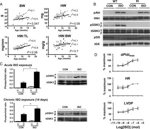
Aims: Glycogen synthase kinase 3 (GSK-3) signalling is implicated in the growth of the heart during development and in response to stress. However, its precise role remains unclear. We set out to characterize developmental growth and response to chronic isoproterenol (ISO) stress in knockin (KI) mice lacking the critical N-terminal serines, 21 of GSK-3alpha and 9 of GSK-3beta respectively, required for inactivation by upstream kinases.
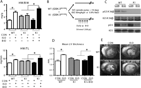
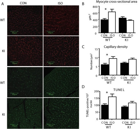
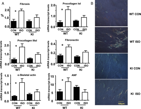
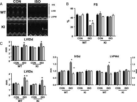
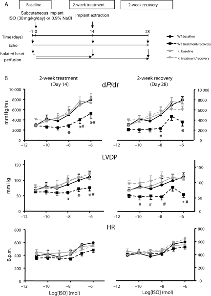
Methods and results: Between 5 and 15 weeks, KI mice grew more rapidly, but normalized heart weight and contractile performance were similar to wild-type (WT) mice. Isolated hearts of both genotypes responded comparably to acute ISO infusion with increases in heart rate and contractility. In WT mice, chronic subcutaneous ISO infusion over 14 days resulted in cardiac hypertrophy, interstitial fibrosis, and impaired contractility, accompanied by foetal gene reactivation. These effects were all significantly attenuated in KI mice. Indeed, ISO-treated KI hearts demonstrated reversible physiological remodelling traits with increased stroke volume and a preserved contractile response to acute adrenergic stimulation. Furthermore, simultaneous pharmacological inhibition of GSK-3 in KI mice treated with chronic subcutaneous ISO recapitulated the adverse remodelling phenotype seen in WT hearts.
Conclusion: Expression of inactivation-resistant GSK-3alpha/beta does not affect eutrophic myocardial growth but protects against pathological hypertrophy induced by chronic adrenergic stimulation, maintaining cardiac function and attenuating interstitial fibrosis. Accordingly, strategies to prevent phosphorylation of Ser-21/9, and consequent inactivation of GSK-3alpha/beta, may enable a sustained cardiac response to chronic beta-agonist stimulation while preventing pathological remodelling.
Figures
Similar articles
-
Myocardial stress remodelling after regional infarction is independent of glycogen synthase kinase-3 inactivation.J Mol Cell Cardiol. 2010 Nov;49(5):897-900. doi: 10.1016/j.yjmcc.2010.07.021. Epub 2010 Aug 6. J Mol Cell Cardiol. 2010. PMID: 20696171 Free PMC article.
-
Phosphorylation of eukaryotic translation initiation factor 2Bepsilon by glycogen synthase kinase-3beta regulates beta-adrenergic cardiac myocyte hypertrophy.Circ Res. 2004 Apr 16;94(7):926-35. doi: 10.1161/01.RES.0000124977.59827.80. Epub 2004 Mar 4. Circ Res. 2004. PMID: 15001529
-
Glycogen synthase kinase-3alpha reduces cardiac growth and pressure overload-induced cardiac hypertrophy by inhibition of extracellular signal-regulated kinases.J Biol Chem. 2007 Nov 9;282(45):33181-91. doi: 10.1074/jbc.M705133200. Epub 2007 Sep 12. J Biol Chem. 2007. PMID: 17855351
-
Role of GSK-3 in Cardiac Health: Focusing on Cardiac Remodeling and Heart Failure.Curr Drug Targets. 2021;22(13):1568-1576. doi: 10.2174/1389450122666210224105430. Curr Drug Targets. 2021. PMID: 33655828 Review.
-
Glycogen synthase kinase 3 (GSK3) in the heart: a point of integration in hypertrophic signalling and a therapeutic target? A critical analysis.Br J Pharmacol. 2008 Mar;153 Suppl 1(Suppl 1):S137-53. doi: 10.1038/sj.bjp.0707659. Epub 2008 Jan 21. Br J Pharmacol. 2008. PMID: 18204489 Free PMC article. Review.
Cited by
-
2,5-Dimethylcelecoxib prevents pressure-induced left ventricular remodeling through GSK-3 activation.Hypertens Res. 2017 Feb;40(2):130-139. doi: 10.1038/hr.2016.122. Epub 2016 Sep 15. Hypertens Res. 2017. PMID: 27628899
-
A rat model of metabolic syndrome-related heart failure with preserved ejection fraction phenotype: pathological alterations and possible molecular mechanisms.Front Cardiovasc Med. 2023 Jul 4;10:1208370. doi: 10.3389/fcvm.2023.1208370. eCollection 2023. Front Cardiovasc Med. 2023. PMID: 37469482 Free PMC article.
-
Enhanced catecholamine release in mice expressing PKB/SGK-resistant GSK3.Pflugers Arch. 2011 Dec;462(6):811-9. doi: 10.1007/s00424-011-1006-6. Epub 2011 Sep 16. Pflugers Arch. 2011. PMID: 21922189
-
Hydrogen (H2) Inhibits Isoproterenol-Induced Cardiac Hypertrophy via Antioxidative Pathways.Front Pharmacol. 2016 Oct 27;7:392. doi: 10.3389/fphar.2016.00392. eCollection 2016. Front Pharmacol. 2016. PMID: 27833552 Free PMC article.
-
Kvβ1.1 (AKR6A8) senses pyridine nucleotide changes in the mouse heart and modulates cardiac electrical activity.Am J Physiol Heart Circ Physiol. 2017 Mar 1;312(3):H571-H583. doi: 10.1152/ajpheart.00281.2016. Epub 2016 Dec 16. Am J Physiol Heart Circ Physiol. 2017. PMID: 27986658 Free PMC article.
References
-
- Tong H, Imahashi K, Steenbergen C, Murphy E. Phosphorylation of glycogen synthase kinase-3beta during preconditioning through a phosphatidylinositol-3-kinase-dependent pathway is cardioprotective. Circ Res. 2002;90:377–379. doi:10.1161/01.RES.0000012567.95445.55. - DOI - PubMed
-
- Antos CL, McKinsey TA, Frey N, Kutschke W, McAnally J, Shelton JM, et al. Activated glycogen synthase-3 beta suppresses cardiac hypertrophy in vivo. Proc Natl Acad Sci USA. 2002;99:907–912. doi:10.1073/pnas.231619298. - DOI - PMC - PubMed
-
- Haq S, Choukroun G, Kang ZB, Ranu H, Matsui T, Rosenzweig A, et al. Glycogen synthase kinase-3beta is a negative regulator of cardiomyocyte hypertrophy. J Cell Biol. 2000;151:117–130. doi:10.1083/jcb.151.1.117. - DOI - PMC - PubMed
-
- Haq S, Choukroun G, Lim H, Tymitz KM, del Monte F, Gwathmey J, et al. Differential activation of signal transduction pathways in human hearts with hypertrophy versus advanced heart failure. Circulation. 2001;103:670–677. - PubMed
Publication types
MeSH terms
Substances
Grants and funding
LinkOut - more resources
Full Text Sources
Other Literature Sources
Molecular Biology Databases
