

Notch2 activation in the embryonic kidney depletes nephron progenitors
- PMID: 20299358
- PMCID: PMC2865736
- DOI: 10.1681/ASN.2009040353
Notch2 activation in the embryonic kidney depletes nephron progenitors
Abstract
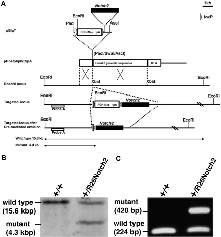
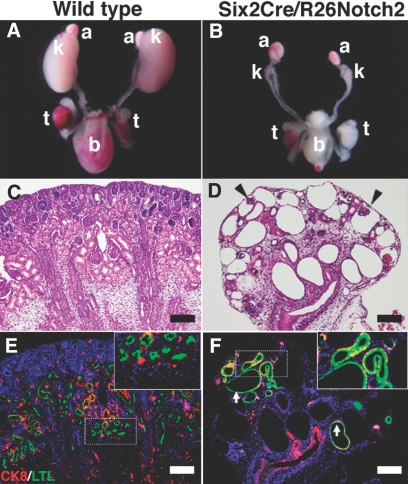
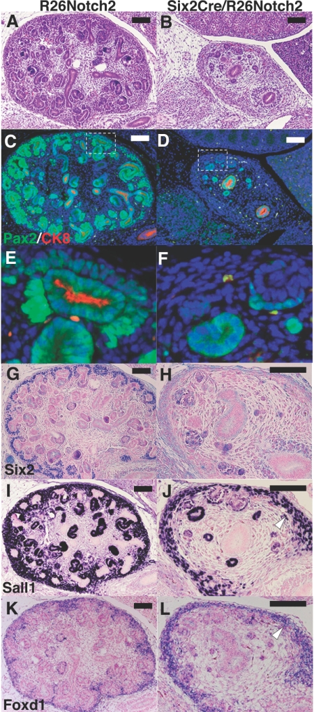
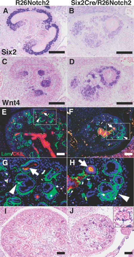
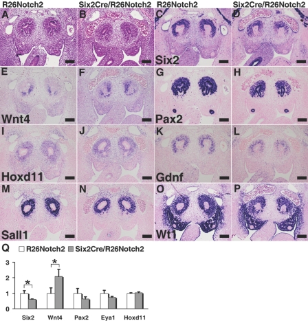
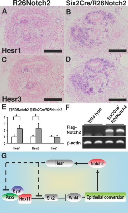
Successive activation of Wnt4 and Notch2 generates nephrons from the metanephric mesenchyme. Mesenchymal-to-epithelial transition requires Wnt4, and normal development of the proximal nephron (epithelia of glomeruli and proximal tubules) requires Notch2. It is unknown, however, whether Notch2 dictates the fate of the proximal nephron directly. Here, we generated a mutant strain of mice with activated Notch2 in Six2-containing nephron progenitor cells of the metanephric mesenchyme. Notch2 activation did not skew the cell fate toward the proximal nephron but resulted in severe kidney dysgenesis and depletion of Six2-positive progenitors. We observed ectopic expression of Wnt4 and premature tubule formation, similar to the phenotype of Six2-deficient mice. Activation of Notch2 in the progenitor cells suppressed Pax2, an upstream regulator of Six2, possibly through Hesr genes. Taken together, these data suggest that a positive feedback loop exists between Notch2 and Wnt4, and that Notch2 stabilizes, rather than dictates, nephron fate by shutting down the maintenance of undifferentiated progenitor cells, thereby depleting this population.
Figures
Similar articles
-
Nephron progenitors in the metanephric mesenchyme.Pediatr Nephrol. 2011 Sep;26(9):1463-7. doi: 10.1007/s00467-011-1806-0. Epub 2011 Feb 19. Pediatr Nephrol. 2011. PMID: 21336811 Review.
-
Stem cells in the embryonic kidney.Kidney Int. 2008 Apr;73(8):913-7. doi: 10.1038/sj.ki.5002784. Epub 2008 Jan 16. Kidney Int. 2008. PMID: 18200005 Review.
-
The contribution of Notch1 to nephron segmentation in the developing kidney is revealed in a sensitized Notch2 background and can be augmented by reducing Mint dosage.Dev Biol. 2010 Jan 15;337(2):386-95. doi: 10.1016/j.ydbio.2009.11.017. Epub 2009 Nov 13. Dev Biol. 2010. PMID: 19914235 Free PMC article.
-
Osr1 acts downstream of and interacts synergistically with Six2 to maintain nephron progenitor cells during kidney organogenesis.Development. 2014 Apr;141(7):1442-52. doi: 10.1242/dev.103283. Epub 2014 Mar 5. Development. 2014. PMID: 24598167 Free PMC article.
-
p53 Enables metabolic fitness and self-renewal of nephron progenitor cells.Development. 2015 Apr 1;142(7):1228-41. doi: 10.1242/dev.111617. Development. 2015. PMID: 25804735 Free PMC article.
Cited by
-
Effect of Hypoxia on Branching Characteristics and Cell Subpopulations during Kidney Organ Culture.Bioengineering (Basel). 2022 Dec 14;9(12):801. doi: 10.3390/bioengineering9120801. Bioengineering (Basel). 2022. PMID: 36551007 Free PMC article.
-
Selective In Vitro Propagation of Nephron Progenitors Derived from Embryos and Pluripotent Stem Cells.Cell Rep. 2016 Apr 26;15(4):801-813. doi: 10.1016/j.celrep.2016.03.076. Epub 2016 Apr 14. Cell Rep. 2016. PMID: 27149838 Free PMC article.
-
The phosphatase Dullard negatively regulates BMP signalling and is essential for nephron maintenance after birth.Nat Commun. 2013;4:1398. doi: 10.1038/ncomms2408. Nat Commun. 2013. PMID: 23360989
-
Developmental Genetics and Congenital Anomalies of the Kidney and Urinary Tract.J Pediatr Genet. 2016 Mar;5(1):51-60. doi: 10.1055/s-0035-1558423. Epub 2015 Sep 7. J Pediatr Genet. 2016. PMID: 27617142 Free PMC article. Review.
-
Notch Signaling in Kidney Development, Maintenance, and Disease.Biomolecules. 2019 Nov 4;9(11):692. doi: 10.3390/biom9110692. Biomolecules. 2019. PMID: 31690016 Free PMC article. Review.
References
-
- Moore MW, Klein RD, Farinas I, Sauer H, Armanini M, Phillips H, Reichardt LF, Ryan AM, Carver-Moore K, Rosenthal A: Renal and neuronal abnormalities in mice lacking GDNF. Nature 382: 76–79, 1996. - PubMed
-
- Pichel JG, Shen L, Sheng HZ, Granholm AC, Drago J, Grinberg A, Lee EJ, Huang SP, Saarma M, Hoffer BJ, Sariola H, Westphal H: Defects in enteric innervation and kidney development in mice lacking GDNF. Nature 382: 73–76, 1996. - PubMed
-
- Sanchez MP, Silos-Santiago I, Frisen J, He B, Lira SA, Barbacid M: Renal agenesis and the absence of enteric neurons in mice lacking GDNF. Nature 382: 70–73, 1996. - PubMed
-
- Carroll TJ, Park JS, Hayashi S, Majumdar A, McMahon AP: Wnt9b plays a central role in the regulation of mesenchymal to epithelial transitions underlying organogenesis of the mammalian urogenital system. Dev Cell 9: 283–292, 2005. - PubMed
-
- Stark K, Vainio S, Vassileva G, McMahon AP: Epithelial transformation of metanephric mesenchyme in the developing kidney regulated by Wnt-4. Nature 372: 679–683, 1994. - PubMed
Publication types
MeSH terms
Substances
LinkOut - more resources
Full Text Sources
Other Literature Sources
Molecular Biology Databases
Miscellaneous
