. 2010 Jun;21(6):943-54.
doi: 10.1681/ASN.2009030242. Epub 2010 Mar 18.

Reactive oxygen species promote caspase-12 expression and tubular apoptosis in diabetic nephropathy

Affiliations

Reactive oxygen species promote caspase-12 expression and tubular apoptosis in diabetic nephropathy

Marie-Luise Brezniceanu et al. J Am Soc Nephrol. 2010 Jun.

Abstract

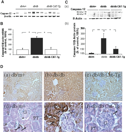
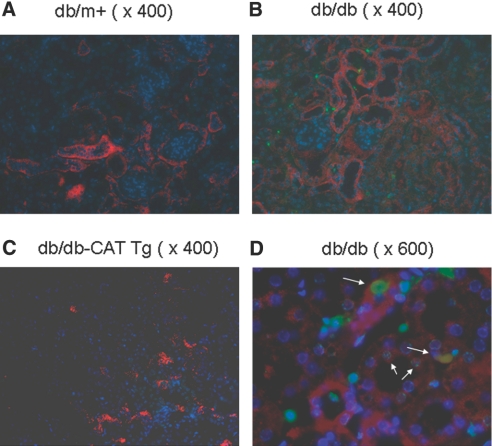
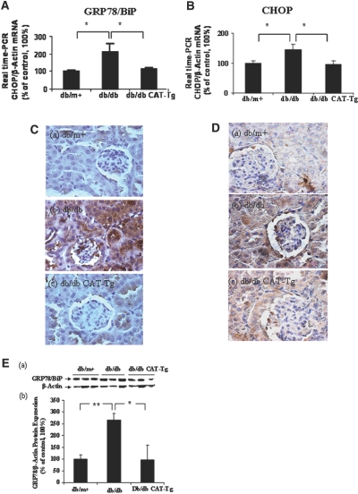
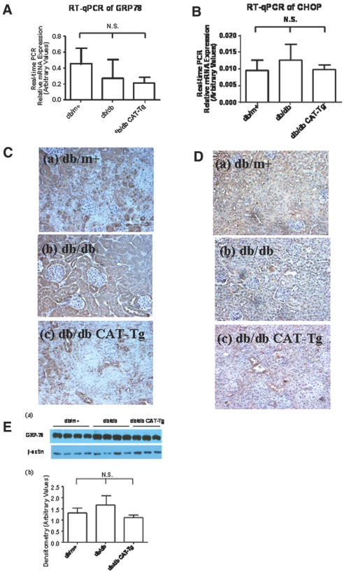
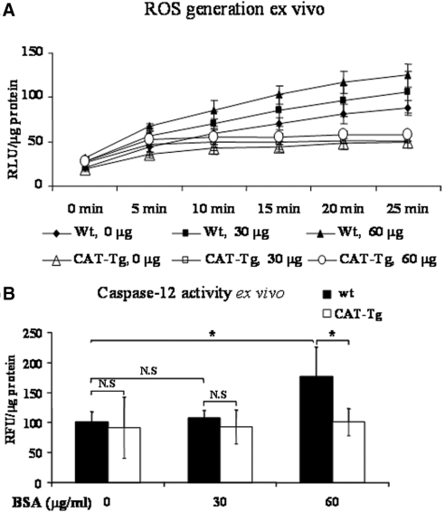
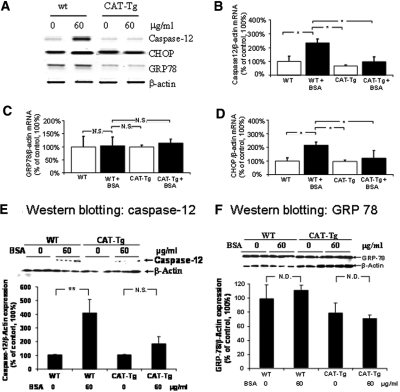
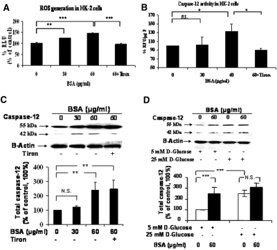
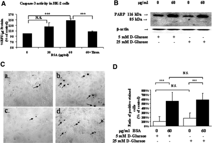
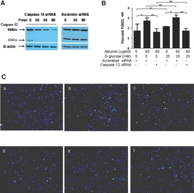
Apoptosis of tubular epithelial cells contributes to the tubular atrophy that accompanies diabetic nephropathy. Reactive oxygen species (ROS) promote tubular apoptosis, but the mechanisms by which this occurs are incompletely understood. Here, we sought proapoptotic genes that ROS differentially upregulate in renal proximal tubular cells of diabetic (db/db) mice. We performed microarray analysis using total RNA from freshly isolated renal proximal tubules of nondiabetic, diabetic, and diabetic transgenic mice overexpressing catalase in the proximal tubule (thereby attenuating ROS). We observed greater expression of caspase-12 in the proximal tubules of the diabetic mice compared with the nondiabetic and diabetic transgenic mice. Quantitative PCR and immunohistochemistry confirmed the enhanced expression of caspase-12, as well as members of the endoplasmic reticulum stress-induced apoptotic pathway. Ex vivo, albumin induced caspase-12 activity and expression (protein and mRNA) and mRNA expression of the CCAT/enhancer-binding protein homologous protein in freshly isolated wild-type proximal tubules but not in catalase-overexpressing proximal tubules. In vitro, albumin stimulated activity of both caspase-12 and caspase-3 as well as expression of caspase-12 and CCAT/enhancer-binding protein homologous protein in a human proximal tubule cell line (HK-2). The free radical scavenger tiron inhibited these effects. Furthermore, knockdown of caspase-12 with small interfering RNA reduced albumin-induced apoptosis in HK-2 cells. Taken together, these studies demonstrate that albuminuria may induce tubular apoptosis through generation of ROS and the subsequent expression and activation of endoplasmic reticulum stress genes in the diabetic kidney.


Figures

Figure 1.
Figure 1.
Caspase-12 expression is validated in mouse kidneys at week 20. (A) Southern blotting of conventional RT-PCR analysis of caspase-12 mRNA expression in RPTs of db/m+, db/db, and db/db CAT-Tg mice. (B) RT-qPCR analysis of mouse caspase-12 mRNA expression in RPTs of db/m+ (wt), db/db, and db/db CAT-Tg mice. Caspase-12 mRNA levels in db/m+ mice were considered as 100%. Each point represents the mean ± SD of six animals. *P < 0.05; **P < 0.01. (C, a) Immunoblotting (antibody from Cell Signaling) of mouse caspase-12 protein expression in RPTs of db/m+ (wt), db/db (db), and db/db CAT-Tg (db-Cat) mice. (b) Densitometry of the data in a. (D) Immunohistochemical staining for caspase-12 in db/m+, db/db, and db/db CAT-Tg mouse kidneys, using rabbit anti–caspase-12 polyclonal antibodies (antibody from eBioscience). Arrows indicate caspase-12 immunopositive cells in proximal tubules. Magnifications: ×200 in D, a through c; ×600 in D, d through f.
Figure 2.
Figure 2.
Caspase-12 expression and apoptotic cells are localized in mouse kidneys at week 20. Twenty-week-old mouse kidneys were sectioned and subjected to TUNEL assay to visualize apoptotic cells (green) and then incubated with anti–caspase-12 antibody (eBioscience) followed by anti-rabbit AlexaFluor 555 to demonstrate caspase-12 expression (red). Cells staining positively for TUNEL and caspase-12 appear yellow. Nuclei were stained with DAPI (blue). (a through c) Kidneys from db/m+ mouse kidney (a), db/db mouse kidney (b), and db/db-CAT Tg (c) mice. Kidneys were first stained with TUNEL and then incubated with anti–caspase-12 antibody. (d) higher magnification of b. Arrows indicate cells that stained positively for TUNEL and caspase-12. Magnification, ×400 in a through c; ×600 in d.
Figure 3.
Figure 3.
GRP78/BiP and CHOP are expressed in mouse kidneys at week 20. (A and B) RPTs from mouse kidneys were isolated and assayed by RT-qPCR for GRP78/BiP (A) and CHOP (B) mRNA. The relative densities of GRP78/BiP and CHOP mRNA were normalized with β-actin mRNA control. GRP78/BiP and CHOP mRNA levels in db/m+ mice were considered as 100%. Each point represents the mean ± SD of six animals. *P < 0.05. (C and D) Immunostaining for GRP78/BiP (C) and CHOP (D) expression in mouse kidneys at 20 weeks. (E, a) Immunoblotting of mouse GRP78/BiP protein expression in RPTs of db/m+, db/db, and db/db CAT-Tg mice. (b) Densitometry of the data in a. Each bar represents the mean ± SD of at least three animals. *P < 0.05; **P < 0.01. Magnification, ×600 in C and D.
Figure 4.
Figure 4.
Caspase-12 is expressed in 12-week-old mouse kidneys. (A) Immunoblotting of caspase-12 protein expression. Each lane represents a different mouse. (B) Densitometry of the data in A. (C) RT-qPCR data showing caspase-12 mRNA expression from db/m+, db/db, and db/db CAT-Tg mice. Each bar represents the mean ± SD of at least three animals. (D) Immunostaining of caspase-12 expression. Magnification, ×200 in D.
Figure 5.
Figure 5.
GRP-78/BiP and CHOP are expressed in 12-week-old mouse kidneys. (A and B) RT-qPCR data showing GRP78/BiP (A) and CHOP (B) mRNA expression from db/m+, db/db, and db/db CAT-Tg mice. Each bar represents the mean ± SD of at least three animals. (C and D) Immunostaining of GRP78/BiP (C) and CHOP (D) expression in mouse kidneys. (E, a) Immunoblotting of GRP78/BiP protein expression. Each lane represents a different mouse. (b) Densitometry of the data in a. Magnification, ×200 in C and D.
Figure 6.
Figure 6.
Albumin effects ROS production and caspase-12 activity in mRPTs ex vivo. mRPTs from male wt or CAT-Tg mice were incubated in 5 mM d-glucose serum-free DMEM in the absence or presence of albumin (0, 30, or 60 μg/ml) for 16 hours. (A) ROS generation was assessed after 10 minutes of incubation in Krebs buffer and expressed as relative light units (RLU). (B) Caspase-12 activity assays were performed after 16 hours of incubation in mRPTs.
Figure 7.
Figure 7.
Albumin affects caspase-12, GRP78/BiP, and CHOP expression in RPTs of wt and CAT-Tg mice ex vivo. (A) Southern blotting of RT-PCR of caspase-12, CHOP, and GRP78/Bip mRNA. (B) RT-qPCR of caspase-12 mRNA. (C) RT-qPCR of GRP78/BiP mRNA. (D) RT-qPCR of CHOP mRNA. (E, a) Immunoblotting of caspase-12 protein expression. (b) Densitometry of the data in a. (F, a) immunoblotting of GRP-78/BiP protein expression. (b) Densitometry of the immunoblotting of a.
Figure 8.
Figure 8.
Albumin affects ROS production, caspase-12 activity, and caspase-12 expression in HK-2 cells in vitro. HK-2 cells were incubated in 5 mM d-glucose serum-free DMEM in the absence or presence of albumin (0, 30, or 60 μg/ml) with or without tiron (10−4 M) for 16 hours. (A) ROS generation. (B) Caspase-12 activity assay. (C) Caspase-12 protein expression assessed by immunoblotting and quantification. (D) Immunoblotting and quantification of caspase-12 protein expression in HK-2 cells in 5 mM d-glucose plus 20 mM d-mannitol or 25 mM d-glucose in the presence or absence of 60 μg/ml albumin. *P < 0.05; **P < 0.01; ***P < 0.005.
Figure 9.
Figure 9.
Albumin affects caspase-3 activity and apoptosis in HK-2 cells in vitro. HK-2 cells were incubated in 5 mM d-glucose serum-free DMEM in the absence or presence of albumin (0, 30, or 60 μg/ml) with or without tiron (10−4 M) for 16 hours. (A) Caspase-3 activity assay. (B) Immunoblotting for PARP expression and cleavage in HK-2 cells in 5 mM d-glucose plus 20 mM d-mannitol or 25 mM d-glucose in the presence or absence of 60 μg/ml albumin. (C) HK-2 cell apoptosis analyzed by TUNEL staining assay. HK-2 cells were incubated in 5 mM d-glucose plus 20 mM d-mannitol medium in the absence (a) or in the presence (b) of 60 μg/ml albumin or in 25 mM d-glucose medium in the absence (c) or presence (d) of 60 μg/ml albumin. Arrows indicate the TUNEL-positive cells. (D) Quantification of TUNEL-positive cells in C. Data are means ± SD; n = 3. ***P < 0.005. Magnification, ×200 in C.
Figure 10.
Figure 10.
Caspase-12 siRNA affects the number of TUNEL-positive apoptotic cells. (A) HK-2 cells were incubated in 5 mM d-glucose medium with increasing dosages of either caspase-12 siRNA or scrambled siRNA. (B) HK-2 cells were incubated in normal-glucose (5 mM d-glucose plus 20 mM d-mannitol) or high-glucose (25 mM d-glucose) medium in the absence or presence of albumin (60 μg/ml) and with or without caspase-12 siRNA or scrambled siRNA (40 pmol). Quantification of TUNEL-positive stained cells. Data are means ± SD. *P < 0.05; **P < 0.01. (C) Apoptosis was assessed by the TUNEL assay. TUNEL-positive apoptotic cells fluoresced green, and DAPI-stained nuclei fluoresced blue. HK-2 cells, plated at a density of 2.5 × 10⁴ in 12-well microplates, were incubated in 5 mM d-glucose plus 20 mM d-mannitol in the absence of albumin and presence of 40 pmol of scrambled siRNA (a) or in the presence of 60 μg/ml albumin and 40 pmol of scrambled siRNA (b) or in the presence of 60 μg/ml albumin and 40 pmol of caspase-12 siRNA (c). HK-2 cells were incubated in 25 mM d-glucose medium in the absence of albumin and presence of 40 pmol of scrambled siRNA (d) or in the presence of 60 μg/ml albumin and 40 pmol of scrambled siRNA (e) or in the presence of 60 μg/ml albumin and 40 pmol of caspase-12 siRNA (f). Magnification, ×100 in C.

Comment in

Similar articles

Cited by

References

    1. Rabkin R: Diabetic nephropathy. Clin Cornerstone 5: 1–11, 2003. - PubMed
    1. Sarafidis PA, Stafylas PC, Kanaki AI, Lasaridis AN: Effects of renin-angiotensin system blockers on renal outcomes and all-cause mortality in patients with diabetic nephropathy: An updated meta-analysis. Am J Hypertens 21: 922–929, 2008. - PubMed
    1. Gordois A, Scuffham P, Shearer A, Oglesby A: The health care costs of diabetic nephropathy in the United States and the United Kingdom. J Diabetes Complications 18: 18–26, 2004. - PubMed
    1. Hovind P, Tarnow L, Parving HH: Remission and regression of diabetic nephropathy. Curr Hypertens Rep 6: 377–382, 2004. - PubMed
    1. Tarchini R, Bottini E, Botti P, Marseglia CD, Talassi E, Baraldi O, Lambertini D, Gaetti L, Bellomi A: Type 2 diabetic nephropathy: Clinical course and prevention proposals 2004 [in Italian]. G Ital Nefrol 22[Suppl 31]: S15–S19, 2005. - PubMed

Publication types

MeSH terms