

Wallerian degeneration in the corticospinal tract evaluated by diffusion tensor imaging correlates with motor deficit 30 days after middle cerebral artery ischemic stroke
- PMID: 20299434
- PMCID: PMC7965455
- DOI: 10.3174/ajnr.A2038
Wallerian degeneration in the corticospinal tract evaluated by diffusion tensor imaging correlates with motor deficit 30 days after middle cerebral artery ischemic stroke
Abstract
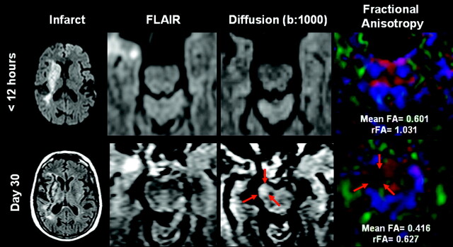
Background and purpose: The quantification and clinical significance of WD in CSTs following supratentorial stroke are not well understood. We evaluated the anisotropy by using DTI and signal-intensity changes on conventional MR imaging in the CST to determine whether these findings are correlated with limb motor deficit in patients with MCA ischemic stroke.
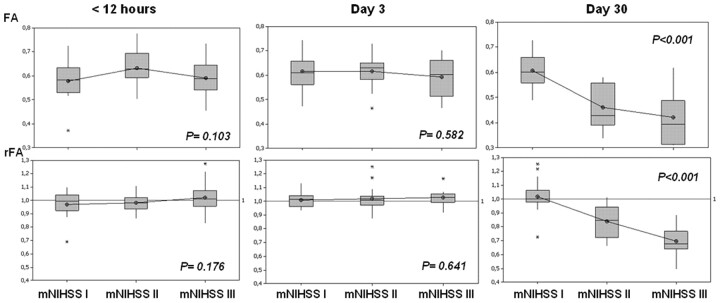
Materials and methods: We studied 60 patients within 12 hours of stroke onset. At admission, day 3, and day 30 of evolution, patients underwent multimodal MR imaging, including DTI sequences. We assessed the severity of limb weakness by using the motor subindex scores (5a, 5b, 6a, 6b) of the m-NIHSS and established 3 groups: I (m-NIHSS scores of 0), II (m-NIHSS, 1-4), and III (m-NIHSS, 5-8). FA values and rFAs were measured on the affected and the unaffected CSTs in the pons.
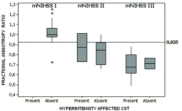
Results: FA values for the CST were significantly lower on the affected side compared with the unaffected side only at day 30 (P < .001), and the rFA was significantly correlated with the motor deficit at day 30 (P < .001; r = -0.793). The sensitivity, specificity, and positive and negative predictive values for motor deficit by rFA < 0.925 were 95.2%, 94.9%, 90.9%, and 97.4%, respectively.
Conclusions: WD in the CST revealed by DTI correlates with motor deficit 30 days after MCA ischemic stroke. This study highlights the utility of imaging follow-up at 30 days and the potential of DTI as a surrogate marker in clinical trials.
Figures
References
-
- Duncan PW, Goldstein LB, Matchar D, et al. . Measurement of motor recovery after stroke: outcome assessment and sample size requirements. Stroke 1992;23:1084–89 - PubMed
-
- Davidoff RA. The corticospinal tract. Neurology 1990;40:332–39 - PubMed
-
- Lindberg PG, Skejø PH, Rounis E, et al. . Wallerian degeneration of the corticofugal tracts in chronic stroke: a pilot study relating diffusion tensor imaging, transcranial magnetic stimulation, and hand function. Neurorehabil Neural Repair 2007;21:551–60 - PubMed
-
- Iizuka H, Sakatani K, Young W. Corticofugal axonal degeneration in rats after middle cerebral artery occlusion. Stroke 1989;20:1396–402 - PubMed
Publication types
MeSH terms
LinkOut - more resources
Full Text Sources
Medical