

Syndromes of the first and second branchial arches, part 1: embryology and characteristic defects
- PMID: 20299437
- PMCID: PMC7964959
- DOI: 10.3174/ajnr.A2072
Syndromes of the first and second branchial arches, part 1: embryology and characteristic defects
Abstract
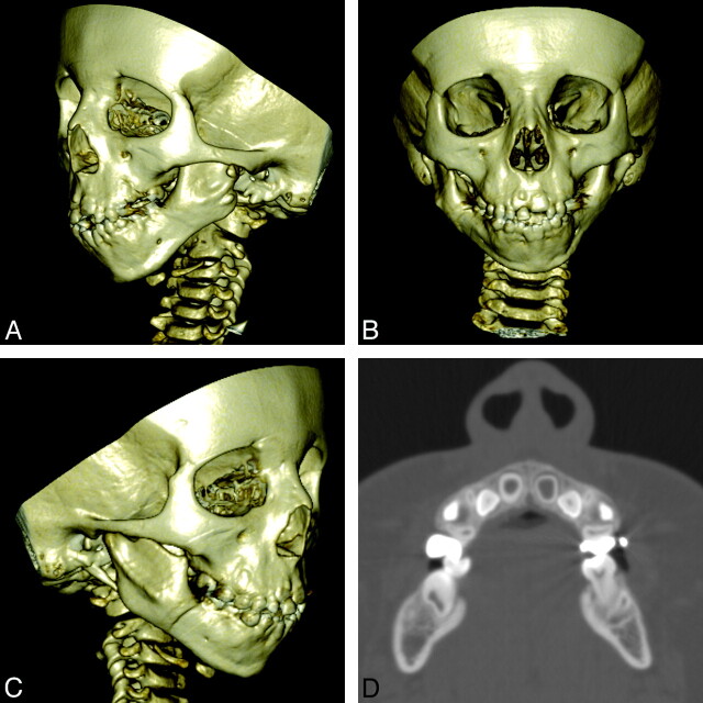
A variety of congenital syndromes affecting the face occur due to defects involving the first and second BAs. Radiographic evaluation of craniofacial deformities is necessary to define aberrant anatomy, plan surgical procedures, and evaluate the effects of craniofacial growth and surgical reconstructions. High-resolution CT has proved vital in determining the nature and extent of these syndromes. The radiologic evaluation of syndromes of the first and second BAs should begin first by studying a series of isolated defects: CL with or without CP, micrognathia, and EAC atresia, which compose the major features of these syndromes and allow more specific diagnosis. After discussion of these defects and the associated embryology, we proceed to discuss the VCFS, PRS, ACS, TCS, Stickler syndrome, and HFM.
Figures

References
-
- Marsh JL. Comprehensive Care for Craniofacial Deformities. St. Louis: Mosby; 1985
-
- Passos-Bueno MR, Ornelas CC, Fanganiello RD. Syndromes of the first and second pharyngeal arches: a review. Am J Med Genet A 2009;149A:1853–59 - PubMed
-
- Walker MB, Trainor PA. Craniofacial malformations: intrinsic vs extrinsic neural crest cell defects in Treacher Collins and 22q11 deletion syndromes. Clin Genet 2006;69:471–79 - PubMed
-
- Moraes F, Novoa A, Jerome-Majewska LA, et al. . Tbx1 is required for proper neural crest migration and to stabilize spatial patterns during middle and inner ear development. Mech Dev 2005;122:199–212 - PubMed
Publication types
MeSH terms
LinkOut - more resources
Full Text Sources
Medical
Miscellaneous