. 2010 May 14;285(20):15627-15636.
doi: 10.1074/jbc.M109.079483. Epub 2010 Mar 18.

Dicalcin inhibits fertilization through its binding to a glycoprotein in the egg envelope in Xenopus laevis

Affiliations

Dicalcin inhibits fertilization through its binding to a glycoprotein in the egg envelope in Xenopus laevis

Naofumi Miwa et al. J Biol Chem. .

Abstract

Fertilization comprises oligosaccharide-mediated sperm-egg interactions, including sperm binding to an extracellular egg envelope, sperm penetration through the envelope, and fusion with an egg plasma membrane. We show that Xenopus dicalcin, an S100-like Ca(2+)-binding protein, present in the extracellular egg envelope (vitelline envelope (VE)), is a suppressive mediator of sperm-egg interaction. Preincubation with specific antibody greatly increased the efficiency of in vitro fertilization, whereas prior application of exogenous dicalcin substantially inhibited fertilization as well as sperm binding to an egg and in vitro sperm penetration through the VE protein layer. Dicalcin showed binding to protein cores of gp41 and gp37, constituents of VE, in a Ca(2+)-dependent manner and increased in vivo reactivity of VE with a lectin, Ricinus communis agglutinin I, which was accounted for by increased binding ability of gp41 to the lectin and greater exposure of gp41 to an external environment. Our findings strongly suggest that dicalcin regulates the distribution of oligosaccharides within the VE through its binding to the protein core of gp41, probably by modulating configuration of oligosaccharides on gp41 and the three-dimensional structure of VE framework, and thereby plays a pivotal role in sperm-egg interactions during fertilization.


Figures

FIGURE 1.
FIGURE 1.
Ca2+ binding activity of Xenopus dicalcin. A, 45Ca blot of Xenopus dicalcin. Recombinant dicalcin and molecular size markers (∼6 μg each) were electrophoresed and transferred onto a PVDF membrane. Blots of recombinant Xenopus dicalcin and size markers were soaked in Tris-buffered saline containing 1 mm 45CaCl2. After washing, the membrane was dried, and bound 45Ca was detected. CBB, Coomassie brilliant blue staining; 45Ca, 45Ca blot; Marker, molecular size markers; Dicalcin, recombinant Xenopus dicalcin. B, Ca2+ binding to Xenopus dicalcin. Recombinant dicalcin (final concentration, 10 μm) was incubated with various concentrations of 45CaCl2. The graph shows the amount of Ca2+ bound to Xenopus dicalcin as a function of free Ca2+ concentration (n = 6, mean ± S.D.). The data were fitted by a Hill equation (solid line).
FIGURE 2.
FIGURE 2.
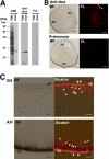
Localization of dicalcin in Xenopus eggs. A, specificity of anti-dicalcin antibody. Left (CBB): CBB staining of the proteins after SDS-PAGE of the homogenate of Xenopus eggs (Egg) and recombinant dicalcin (Dica). Middle (Anti-dica): Western blot analysis of homogenates of Xenopus eggs (Egg) and recombinant dicalcin (Dica) treated with anti-dicalcin antibody. Right (Pre): Blots treated with preimmune antibody. B, immunohistochemical staining at a low magnification. Eggs were fixed, and serial sections (∼14-μm thickness) were treated with anti-dicalcin antibody (Anti-dica) or preimmune antibody (Preimmune). Dicalcin is localized in the marginal area of Xenopus eggs (arrows). BF, bright field observation; FL, fluorescence image. Anti-dica, stained with anti-dicalcin antibody; Preimmune, stained with preimmune antibody. Scale bar: 200 μm. C, detailed localization of dicalcin in Xenopus eggs. Dicalcin is localized in the extracellular vitelline envelope (arrowheads) and in the cytosolic cortex of the egg (arrows). BF, bright field observation; Dicalcin, merged image of bright field and fluorescence image stained with anti-dicalcin antibody. VH, vegetal hemisphere; AH, animal hemisphere; and VE, vitelline envelope. Scale bar: 20 μm.
FIGURE 3.
FIGURE 3.
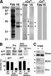
Ca2+-dependent binding of dicalcin to protein cores of gp41 and gp37. A, dicalcin binds to several egg and VE proteins. Soluble egg proteins and vitelline envelope (VE) proteins were prepared as stated under “Experimental Procedures.” VE proteins are composed of four major proteins (asterisks). Blots of both egg-soluble proteins and VE proteins were probed by biotinylated dicalcin either in the presence or absence of Ca2+. Dicalcin bound to several egg proteins and two VE proteins (gp41 and gp37) in the presence of Ca2+ (arrowheads), but not in the absence of Ca2+. Egg, soluble egg proteins; VE, VE proteins. +Ca2+, blot overlay analysis in the presence of Ca2+ (500 μm CaCl2); −Ca2+, analysis in the absence of Ca2+ (500 μm EGTA). B, anti-dicalcin antibody inhibits the binding of dicalcin to gp41 and gp37. Blots of VE proteins were probed with biotinylated dicalcin either in the presence of anti-dicalcin antibody (Anti-dicalcin; 5 and 50 mg/liter) or preimmune antibody (Pre; 50 mg/liter). Silver, silver-stained VE proteins after SDS-PAGE; Pre, blot overlay analysis treated with preimmune antibody; Anti-dicalcin, blot overlay analysis treated with anti-dicalcin antibody. The graph shows mean data (n = 6; *, p = 0.0015; **, p = 2.4 × 10⁻⁵). C, dicalcin binds to deglycosylated forms of gp41 and gp37. VE proteins were treated with TFMS for 3 h. Blots of glycosylated and deglycosylated proteins were probed either with fluorescently labeled RCAI or biotinylated dicalcin. Silver, silver-stained proteins treated with TFMS (TFMS +) or without treatment (TFMS −); RCAI, blot with RCAI; Dica, blot with biotinylated dicalcin. Arrowheads indicate the positions of gp41 and gp37.
FIGURE 4.
FIGURE 4.
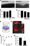
Inhibitory effect of dicalcin on sperm binding and sperm penetration through the VE protein layer. A, representative micrographs of unfertilized (Unfertilized) and fertilized (Fertilized) eggs after sperm treatment. Scale bar: 20 μm. B, effect of pretreatment with anti-dicalcin antibody on sperm binding to egg. Ovulated eggs were pretreated with preimmune antibody (Pre) or anti-dicalcin antibody (Anti-dica) followed by insemination. After rinse, bound sperm at an equatorial plane were counted (n = 9; *, p = 0.027). Almost no sperm remained attached to fertilized eggs (Fertilized). C, effect of pretreatment with dicalcin on sperm binding to egg. Ovulated eggs were pretreated with BSA or dicalcin at the indicated concentrations, and bound sperm were counted (n = 9; *, p = 0.003). D, a scheme that represents our in vitro penetration assay. Suspension of viscous VE proteins (VE) was placed on the mesh of the upper chamber. Some sperm (S) successfully penetrated through the VE. E, representative confocal image of Rhodamine-RCAI-treated VE proteins on the mesh of the upper chamber (red square in D). VE proteins stained with Rhodamine-RCAI (red) were observed. A penetrating spermatozoon stained with Hoechst 33342 was recognized in XZ and YZ planes (arrows). The potential trait of the spermatozoon through the VE is indicated (arrowheads). A direction from the upper chamber to the lower one is indicated (arrows at upper right). m, mesh. Scale bar: 50 μm. F, time course of sperm penetration. Sperm in the lower chamber were collected at the indicated times, and the percentage of the number of the penetrated sperm was calculated. The extent of penetration reached maximum at around 10 min. G, effect of pretreatment with dicalcin on sperm penetration. VE proteins on the upper chamber were pretreated either with dicalcin (4 μm) or BSA (4 μm) for 15 min, followed by sperm placement. After 5 min, sperm in the lower chamber were collected and the number of the penetrated sperm was calculated. The extent of penetration pretreated with BSA was set to ∼100% (n = 6; *, p = 0.039). BSA, pretreated with BSA; dicalcin, pretreated with dicalcin.
FIGURE 5.
FIGURE 5.
Inhibitory effect of dicalcin on in vitro fertilization. A, effect of pretreatment with anti-dicalcin antibody on fertilization. Ovulated eggs were pretreated with preimmune antibody (Pre, 50 mg/liter) or anti-dicalcin (Anti-dica, 5 and 50 mg/liter) followed by incubation of sperm. The number of two-cell embryos was counted until one of the embryos proceeded to the four-cell-stage. Fertilization success was scored and normalized (n = 6; *, p = 0.007). B, effect of pretreatment with dicalcin on fertilization. Ovulated eggs were pretreated with BSA or dicalcin at indicated concentrations followed by insemination (n = 6; *, p = 0.004; **, p = 7.2 × 10⁻⁵). EGTA: addition of EGTA (16 μm) prior to inseminations. +Anti-dica: addition of anti-dicalcin antibody (50 mg/liter) together with dicalcin. C, effect of preincubation of sperm with dicalcin on fertilization. Ovulated eggs were inseminated with sperm (SP) that were preincubated with dicalcin (Dica). As a control, eggs were pretreated with BSA (BSA) similarly as described above and inseminated with sperm.
FIGURE 6.
FIGURE 6.
Dicalcin induced increases in RCAI reactivity of vitelline envelope. A, blots of vitelline envelope (VE) were treated with one of fluorescently labeled lectins (RCAI, soybean agglutinin (SBA), or S. nigra (SNA). RCAI recognized gp41 (arrowhead). B, representative confocal images of a Xenopus egg treated with RCAI. Unfertilized eggs were preincubated either with BSA or dicalcin, followed by RCAI staining. Insets: higher magnified images. RCAI signal at the interface between VE and egg plasma membrane is indicated (arrow). Scale bar: 50 μm. C, averaged line scans of RCAI staining across the VE under lower sensitive detection. Intensities of RCAI staining were profiled across the VE (dashed line in B) either in the preincubation of BSA (BSA) or dicalcin (Dica) (n = 7). The position where RCAI signal starts to rise is designated as 0 μm in the x axis. D, averaged line scans of RCAI staining across the VE under higher sensitive detection. Intensities of RCAI staining were profiled as stated in C (n = 7). The dashed line shows the saturating level of the intensity detection. An increase in RCAI signal at the interface between VE and egg plasma membrane is indicated (arrow). E, neither BSA nor dicalcin reacted with RCAI. CBB, CBB-staining of BSA and dicalcin after SDS-PAGE; RCAI, RCAI blot.
FIGURE 7.
FIGURE 7.
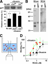
Dicalcin binding to gp41 increases the reactivity of gp41 with RCAI. A, blots of VE proteins were preincubated in the presence or absence of dicalcin and Ca2+, followed by incubation with Rhodamine-labeled RCAI. Representative RCAI binding to gp41 was indicated (upper). The normalized intensity was significantly increased in the presence of dicalcin and Ca2+ (lower graph, n = 8; *, p = 0.06; **, p = 0.002). No add, nothing preincubated; +Dicacin, preincubation with dicalcin (1 μm); +Ca2+, preincubation in the presence of Ca2+ (500 μm CaCl2); −Ca2+, preincubation in the absence of Ca2+ (500 μm EGTA). B, isolation of gp41. Silver, silver-stained VE proteins and isolated gp41; RCAI, blot with RCAI. C, a scheme that represents FCS measurement. A fluorescent signal of diffusing molecule (red) was detected within a small defined confocal volume (yellow) of the well. Autocorrelation analyses of the fluctuating fluorescent signal estimate the diffusion time of fluorescent molecules and distinguish small fast diffusing (i.e. free fluorescent molecules) and large slow moving (target-bound molecules); this enabled us to investigate the stoichiometry of binding. D, the binding of TMR-labeled gp41 to RCAI was analyzed with FCS either in the presence of 1 μm dicalcin (red circles) or absence of dicalcin (black circles) (n = 15). Maximum and minimum diffusion time in each measurement was set to 100 and 0% binding, respectively. Each group of data were fitted with a Hill equation.
FIGURE 8.
FIGURE 8.
Dicalcin binding to gp41 increases exposure of gp41 to an external environment. A, a scheme that represents in vivo labeling of the VE with fluorescent dye, Cy5. Dejellied eggs were labeled with exogenously applied Cy5 in 0.3×MMR. B, after preincubation either with BSA (BSA) or dicalcin (Dica), labeled VE proteins were electrophoresed and fluorescent image was obtained, followed by CBB staining to quantitate the amount of each VE protein (asterisks). CBB, CBB staining of VE proteins; Fluorescent, fluorescent image of Cy5-labeled VE proteins. BSA, preincubated with BSA; Dica, preincubated with dicalcin. C, the ratio of the molar amount of labeled protein to the total amount existing in the preparation was calculated for each VE protein. The highest ratio for gp120, when preincubated with dicalcin, was set to 100% in each trial, and data were normalized. BSA, preincubated with BSA; Dicalcin, preincubated with dicalcin (n = 8–10; *, p < 0.02; **, p = 0.002).
FIGURE 9.
FIGURE 9.
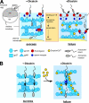
Schematic models of the inhibitory action of dicalcin on fertilization in X. laevis. A, glycoprotein model that involves allosteric conformational change of gp41 caused by dicalcin. In a currently favored model in Xenopus egg, acrosome-intact sperm bind to gp41 (a frog orthologue of mouse ZP3; a major binding partner of sperm) and gp69/64 (an orthologue of mouse ZP2) via carbohydrate moieties (sperm receptors). After acrosome reaction, sperm penetrate through the VE and fuse with an egg (−Dicalcin). The Ca2+-bound form of dicalcin binds to gp41 (and gp37 additionally), leading to a conformational change that could cause an exposure of RCAI ligands. This allosteric change in the configuration of oligosaccharides may mask sperm receptors, forming a functional barrier to prevent sperm binding, penetration, and fusion (+Dicalcin). AH, animal hemisphere; VH, vegetal hemisphere. B, VE structure model that involves structural change in the VE framework caused by dicalcin. VE proteins (gp37, gp41, gp69/64, and gp120) associate with each other and constitute the filament of VE (depicted in −Dicalcin; modified from the schematic model in mammalian ZP). Dicalcin binding to gp37 and gp41 caused a structural change in the three-dimensional structure of the filamentous VE network. This change may involve disorganization of filaments, including fasciculation of filaments, and cause enlargement of the pore size among VE filaments as depicted in +Dicalcin. Symbols are the same in A and B.

Similar articles

Cited by

References

    1. Yanagimachi R. (1994) in The Physiology of Reproduction (Knobil E., Neill J. D. eds) pp. 189–317, Raven Press, New York
    1. Primakoff P., Myles D. G. (2002) Science 296, 2183–2185 - PubMed
    1. Hoodbhoy T., Dean J. (2004) Reproduction 127, 417–422 - PubMed
    1. Litscher E. S., Wassarman P. M. (2007) Histol. Histopathol. 22, 337–347 - PubMed
    1. Clark G. F., Dell A. (2006) J. Biol. Chem. 281, 13853–13856 - PubMed

Publication types