. 2010 Aug;12(8):804-14.
doi: 10.1093/neuonc/noq023. Epub 2010 Mar 18.

Regression of advanced rat and human gliomas by local or systemic treatment with oncolytic parvovirus H-1 in rat models

Affiliations

Regression of advanced rat and human gliomas by local or systemic treatment with oncolytic parvovirus H-1 in rat models

Karsten Geletneky et al. Neuro Oncol. 2010 Aug.

Abstract

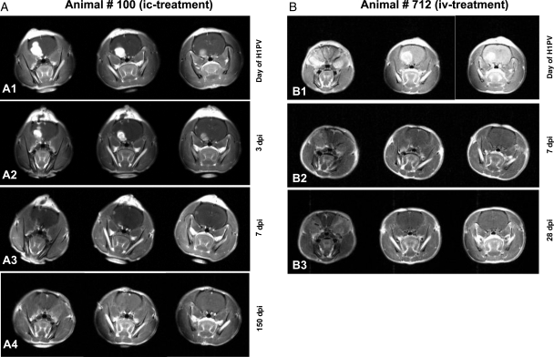
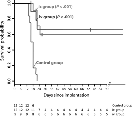
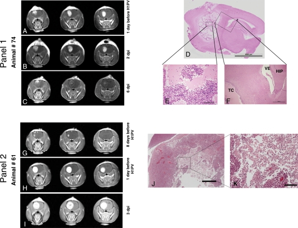
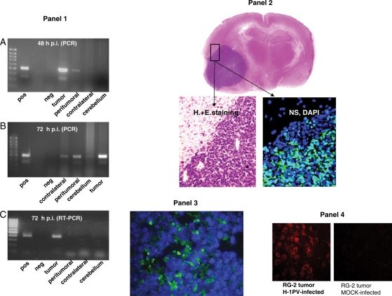
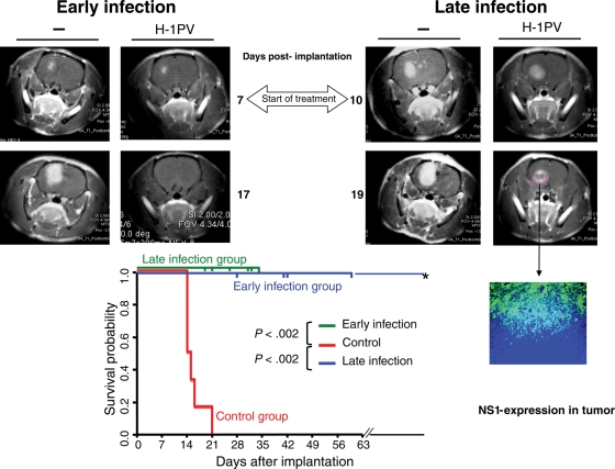
Oncolytic virotherapy is a potential treatment modality under investigation for various malignancies including malignant brain tumors. Unlike some other natural or modified viruses that show oncolytic activity against cerebral neoplasms, the rodent parvovirus H-1 (H-1PV) is completely apathogenic in humans. H-1PV efficiently kills a number of tumor cells without harm to corresponding normal ones. In this study, the concept of H-1PV-based virotherapy of glioma was tested for rat (RG-2 cell-derived) and for human (U87 cell-derived) gliomas in immunocompetent and immunodeficient rat models, respectively. Large orthotopic rat and human glioma cell-derived tumors were treated with either single stereotactic intratumoral or multiple intravenous (iv) H-1PV injections. Oncolysis was monitored by magnetic resonance imaging and proven by histology. Virus distribution and replication were determined in brain and organs. In immunocompetent rats bearing RG-2-derived tumors, a single stereotactic intratumoral injection of H-1PV and multiple systemic (iv) applications of the virus were sufficient for remission of advanced and even symptomatic intracranial gliomas without damaging normal brain tissue or other organs. H-1PV therapy resulted in significantly improved survival (Kaplan-Meier analysis) in both the rat and human glioma models. Virus replication in tumors indicated a contribution of secondary infection by progeny virus to the efficiency of oncolysis. Virus replication was restricted to tumors, although H-1PV DNA could be detected transiently in adjacent or remote normal brain tissue and in noncerebral tissues. The results presented here and the innocuousness of H-1PV for humans argue for the use of H-1PV as a powerful means to perform oncolytic therapy of malignant gliomas.


Figures

Fig. 1.
Fig. 1.
Complete remission of RG-2 gliomas after H-1PV treatment MR images of animal #100 (A1 to A4) and #712 (B1 to B3) at different time points. For each examination, 3 coronary sections are shown. Tumor volumes defined by the area of contrast enhancement progressively decreased after H-1PV injection. Animal #100 was infected with H-1PV intratumorally (A1) and examined on days 3, 7, and 150 p.i. (A2, A3, and A4, respectively). Examination dates for animal #712, which was infected intravenously (B1), were days 7 and 28 p.i. (B2 and B3). Both animals have survived for more than 6 months and without tumor recurrence.
Fig. 2.
Fig. 2.
Prolonged survival following intratumoral or iv H-1PV treatment of RG-2 gliomas. The survival of H-1PV-treated (ic and iv) and control animals is presented using the Kaplan–Meier method. Survival times of treated animals sacrificed for analysis were censored at the date of sacrifice, marked by vertical bars. Values below the plot give the numbers of animals at risk, equivalent to surviving animals on the respective days. The difference between the treated groups and the controls was statistically significant (log rank test: P < .001) both for ic and iv. See also the “Materials and Methods” section.
Fig. 3.
Fig. 3.
Analyses of oncolytic effects in gliomas after H-1PV infection. Panel 1: Regressing tumor (A–C): Tumor regression by MRI (different time points relative to H-1PV infection, 3 coronary sections shown for each examination date). (A) One day before H-1PV administration (15 days after tumor cell implantation), a large tumor was visible in the right frontal lobe. (B) Two days after stereotactic injection of H-1PV, the area of contrast enhancement had slightly increased in volume but displayed a more inhomogeneous appearance (which was associated with an improvement in clinical symptoms). (C) On day 6 p.i., the tumor showed a massive reduction in contrast uptake and an entirely different staining pattern, with a rim-like appearance and only a few areas of remaining enhancement in the central and the most superficial parts of the tumor. The animal was without any clinical symptoms of cerebral or other origin and was sacrificed on this day for further analysis. (D–F) Histological analysis of the area of H-1PV-induced glioma destruction in above animal. (D) In this sagittal section of the entire right hemisphere of the brain (H&E staining; scale bar 5 mm), a large cavity was visible in the location of the initial tumor, whereas surrounding and distant brain tissue had a normal appearance. (E) At a higher (×20) magnification, the tumor cavity appeared to be composed of islands of remaining vital tumor cells embedded in an amorphous eosinophilic mass. Scattered granulocytes, erythrocytes, and plasma cells were detectable, in particular at the periphery of the tumor cell islands. (F) A ×4 magnification of the oblique box in D shows part of the tumor cavity (TC) and adjacent normal structures of the brain such as the ventricle (VE) and the hippocampus (HIP). A few tumor cells were visible underneath the ependyma of the ventricular wall. Scattered lymphocytes and plasma cells were present in the immediate vicinity of the tumor cavity. Otherwise, no significant pathological alterations were detectable within the normal tissue adjacent to the tumor. Panel 2: Progressing tumor in spite of H-1PV therapy (G–I): MR analysis. (G) Eight days before H-1PV administration (10 days after tumor cell implantation), a small tumor began to develop in the right frontal lobe. (H) One day before treatment with H-1PV, the tumor occupied almost the entire right frontal lobe, demonstrating the rapid growth of the glioma. (I) On day 3 p.i., the tumor had further increased in volume. The animal had become symptomatic and was sacrificed on this day. Of note, the uptake of the contrast agent (gadolinium) was less homogeneous in central areas of the tumor injected with H-1PV (I, middle image) compared with the previous time point (H), similar to early changes in regressing tumor (panel 1, B and C). (J and K) Histological analysis. (J) At a low magnification (×2, scale bar 1000 µm), histological appearance of the tumor (H&E staining) showed pale staining of necrotic tissue in the center. (K) At a higher magnification (×10, scale bar 200 µm), oncolysis was characterized by numerous cells with shrunken nuclei (similar to image E, panel 1) as well as necrotic/autolytic ghost cells indicating lytic activity of H-1PV.
Fig. 4.
Fig. 4.
Distribution of viral and cellular markers in the brain of rats following glioma injection with H-1PV. Panel 1: PCR detection of H-1PV DNA (A and B) and RNA (C). The presence of H-1PV DNA in the treated tumor, adjacent brain tissue (peritumoral), contralateral hemisphere, and cerebellum (as indicated) was determined by PCR 48 hours (A) or 72 hours (B) after intratumoral virus injection. The production of H-1PV transcripts at the same locations was tested by RT–PCR 72 hours after intratumoral injection of H-1PV (C). Amplified fragments were detected after agarose gel electrophoresis. The primers used anneal to exon sequences flanking a small intron, resulting in a slightly larger product amplified from DNA compared with intronless cDNA. pos, positive control (H-1PV plasmid DNA); neg, negative control (double-distilled water); far left lanes, size markers (DNA ladder). Panel 2: Immunohistochemical detection of parvoviral NS-1 proteins. Sections were prepared from RG-2 glioma and adjacent (brain) tissues, collected 2 days after intratumoral injection of 1.6 × 10⁷ pfu of H-1PV. The fluorescence signal indicating the presence of NS-1 proteins is restricted to the tumor area and could not be detected in adjacent tissue (DAPI staining of nuclei on the same image). For orientation, H&E staining of the same brain area is shown at low and high magnification. Panel 3: Immunohistochemical detection of parvoviral NS-1 proteins in glioma tissue after iv injection. Sections were prepared 3 days after iv injection of 108 pfu of H-1PV. The green fluorescence demonstrates the parvoviral NS-1 protein scattered throughout the tumor tissue, proving that iv-administered H-1PV reached the brain tumor and is expressed. Panel 4: Impact of H-1PV infection on cathepsin B expression in RG-2 gliomas. An established RG-2 tumor was injected with H-1PV or mock-treated and removed 72 hours later for sectioning and immunohistochemical detection of cathepsin B. Virus infection correlated with a striking activation of cathepsin B, a known effector of H-1PV-induced glioma cell death. No cathepsin B signal was detected in mock infected tumors.
Fig. 5.
Fig. 5.
H-1PV-induced suppression of human gliomas (established from U87 cells) in RNU rats. (Upper panel) Demonstration of tumor regression by MRI (4 representative rat brains; images at different time points relative to tumor cell implantation [105 U87 cells per animal], as indicated). (Left MRI) At day 7 after tumor cell implantation, a small tumor was visible in the right frontal lobe of the 2 brains shown. “Early infection” (combined ic and iv infection with H-1PV) was performed at this day. After 10 days (day 17 post-implantation), this tumor had disappeared in the treated animal (“H-1PV”), whereas in the control animal (–) the tumor area had largely increased. (Right MRI) At day 10 after implantation of U87 cells, a relatively large tumor was identified by MRI in both animals' brains. At this day, “late infection” was performed as described above for “early infection”. In the treated animal, the tumor stopped growing after ic + iv treatment with H-1PV as demonstrated 9 days p.i. (day 19 post-implantation), in contrast to the continuing development of the tumor in the untreated animal (–). Immunohistological examination of the growth-arrested tumor (cryosection of the treated animal's brain) showed expression of the parvoviral NS-1 protein in the necrotic tumor area. (Lower panel) Significantly improved survival of H-1PV-treated animals compared with controls. Early and late infections refer to the time of virus administration relative to tumor development, corresponding to the treatment of small and large tumors, respectively (see main text). All untreated animals had to be sacrificed because of tumor progression, at the latest on day 21 after tumor cell implantation. All treated animals were killed at various times p.i. for analyses of tumors size and histology. All but one of the treated animals, killed on day 19, survived longer than the controls and were sacrificed for analyses on days indicated separately for early and late infection by vertical bars on the two 100% survival probability lines (horizontal lines at 1.0) representing the survival of the 2 treatment groups. One animal (*) survived for more than 8 months.

Similar articles

Cited by

References

    1. Stummer W, Pichlmeier U, Meinel T, Wiestler OD, Zanella F, Reulen HJ. Fluorescence-guided surgery with 5-aminolevulinic acid for resection of malignant glioma: a randomised controlled multicentre phase III trial. Lancet Oncol. 2006;7:392–401. - PubMed
    1. Stupp R, Mason WP, van den Bent MJ, Weller M, Fisher B, Taphoorn MJ, et al. Radiotherapy plus concomitant and adjuvant temozolomide for glioblastoma. N Engl J Med. 2005;352:987–996. - PubMed
    1. Parato KA, Senger D, Forsyth PA, Bell JC. Recent progress in the battle between oncolytic viruses and tumours. Nat Rev Cancer. 2005;5:965–976. - PubMed
    1. Everts B, van der Poel HG. Replication-selective oncolytic viruses in the treatment of cancer. Cancer Gene Ther. 2005;12:141–161. - PubMed
    1. Lun X, Senger DL, Alain T, Oprea A, Parato K, Stojdl D, et al. Effects of intravenously administered recombinant vesicular stomatitis virus (VSV(deltaM51)) on multifocal and invasive gliomas. J Natl Cancer Inst. 2006;98:1546–1557. - PubMed

Publication types