

Review
doi: 10.1038/nrg2752.
Chromatin structure and the inheritance of epigenetic information
Affiliations
- PMID: 20300089
- PMCID: PMC3760772
- DOI: 10.1038/nrg2752
Item in Clipboard
Review
Chromatin structure and the inheritance of epigenetic information
Nat Rev Genet.
2010 Apr.
Abstract
Although it is widely accepted that the regulation of the chromatin landscape is pivotal to conveying the epigenetic program, it is still unclear how a defined chromatin domain is reproduced following DNA replication and transmitted from one cell generation to the next. Here, we review the multiple mechanisms that potentially affect the inheritance of epigenetic information in somatic cells. We consider models of how histones might be recycled following replication, and discuss the importance of positive-feedback loops, long-range gene interactions and the complex network of trans-acting factors in the transmission of chromatin states.
Figures

Characteristics of a Chromatin Domain. Schematic depicting modifications within chromatin that define different chromatin domains. The green dashed lines represent the separation between two adjacent domains. For simplicity, H2A-H2B have been omitted.

Models of histone deposition during replication. Here we illustrate the means by which old and new histones could be deposited following replication. (A) General simplified view of the replication fork. The MCM helicase complex is shown at the fork. The possible interaction of MCM with the histone chaperone ASF1 is shown. The leading strand shows the replication fork, simplified by depicting DNA polymerases ε (Pol), its interaction with PCNA and the interaction of PCNA with ASF1. On the lagging strand, ASF1 and PCNA could interact at three different steps, first during Okazaki fragments replication by the DNA polymerase δ, second during DNA ligation and third with PCNA at the chromatin following replication. Underneath, we present what we believe are important questions that need to be addressed. (B) Random model of histone segregation. In this model, we do not discriminate between dimer or tetramer deposition. The histones segregate randomly between the leading and lagging replicated DNA strands. (C) Semi-conservative model of histone segregation. Left, the scheme is based on the assumption that the H3-H4 tetramer is divided during DNA replication and the parental H3-H4 segregate as dimers onto the newly replicated DNA strands. The parental histones associate with naïve dimers to reconstitute the tetramer. Right, this scheme posits that parental H3-H4 histones segregate as tetramers resulting in the joint deposition of recycled histones and newly deposited naïve histones.
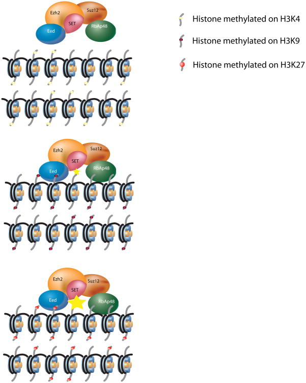
Propagation of H3K27me3 by PRC2. This scheme shows how pre-existing histone methylation marks regulate PRC2 mediated spreading of H3K27 methylation. For simplicity, only one type of histone methylation is presented for each domain although in vivo a combination of them should be taken into consideration. Importantly this scheme does not consider the recruitment of PRC2. Three examples are envisioned: Top, a chromatin domain enriched for an “active mark” such as H3K4me3 that is not recognize by PRC2 and for this reason does not methylate H3K27. Middle, a chromatin domain is enriched for repressive marks such as H3K9me3, H1K26me3 or H4K20me3 that are recognized by PRC2 recognizes but its enzymatic activity is modestly increased (small yellow star). Bottom, a chromatin domain enriched for H3K27me3 that is recognized by PRC2 recognizes this mark and its enzymatic activity is robustly stimulated (large yellow star).

Different phenomena that contribute to propagation of regulatory information. This figure illustrates the different factors that contribute to the propagation of epigenetic information through the regulation of chromatin structure. Regulatory information is represented as a histone posttranslational modification which is diluted by the incorporation of newly synthesized histones during replication. We distinguish two steps: the first one addresses the targeting of the Chromatin Modifying Complex (CMC) at a defined locus, and the second one the spreading of a putative modification throughout a defined domain. Based on known examples, we have attributed defined mechanisms to one step or the other, however it is clear that this distinction is not a strict one and that each of these mechanisms probably contributes to some extent to both steps.
References
-
- Wigler M, Levy D, Perucho M. The somatic replication of DNA methylation. Cell. 1981;24:33–40. - PubMed
-
- Trojer P, Reinberg D. Histone lysine demethylases and their impact on epigenetics. Cell. 2006;125:213–7. - PubMed
-
- Luger K, Mader AW, Richmond RK, Sargent DF, Richmond TJ. Crystal structure of the nucleosome core particle at 2.8 A resolution. Nature. 1997;389:251–60. - PubMed
-
- Kouzarides T. Chromatin modifications and their function. Cell. 2007;128:693–705. - PubMed
Publication types
MeSH terms
Substances
Grants and funding
LinkOut - more resources
Full Text Sources
Other Literature Sources
