The isoforms of the p53 protein
- PMID: 20300206
- PMCID: PMC2829963
- DOI: 10.1101/cshperspect.a000927
The isoforms of the p53 protein
Abstract
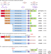
p53 is a transcription factor with a key role in the maintenance of genetic stability and therefore preventing cancer formation. It belongs to a family of genes composed of p53, p63, and p73. The p63 and p73 genes have a dual gene structure with an internal promoter in intron-3 and together with alternative splicing, can express 6 and 29 mRNA variants, respectively. Such a complex expression pattern had not been previously described for the p53 gene, which was not consistent with our understanding of the evolution of the p53 gene family. Consequently, we revisited the human p53 gene structure and established that it encodes nine different p53 protein isoforms because of alternative splicing, alternative promoter usage, and alternative initiation sites of translation. Therefore, the human p53 gene family (p53, p63, and p73) has a dual gene structure. We determined that the dual gene structure is conserved in Drosophila and in zebrafish p53 genes. The conservation through evolution of the dual gene structure suggests that the p53 isoforms play an important role in p53 tumor-suppressor activity. We and others have established that the p53 isoforms can regulate cell-fate outcome in response to stress, by modulating p53 transcriptional activity in a promoter and stress-dependent manner. We have also shown that the p53 isoforms are abnormally expressed in several types of human cancers, suggesting that they play an important role in cancer formation. The determination of p53 isoforms' expression may help to link clinical outcome to p53 status and to improve cancer patient treatment.
Figures


References
-
- Anensen N, Oyan AM, Bourdon JC, Kalland KH, Bruserud O, Gjertsen BT 2006. A distinct p53 protein isoform signature reflects the onset of induction chemotherapy for acute myeloid leukemia. Clin Cancer Res 12:3985–3992 - PubMed
-
- Avery-Kiejda KA, Zhang XD, Adams LJ, Scott RJ, Vojtesek B, Lane DP, Hersey P 2008. Small molecular weight variants of p53 are expressed in human melanoma cells and are induced by the DNA-damaging agent cisplatin. Clin Cancer Res 14:1659–1668 Epub 2008 Feb 1629 - PubMed
-
- Bénard J, Douc-Rasy S, Ahomadegbe JC 2003. TP53 family members and human cancers. Hum Mutat 21:182–191 - PubMed
-
- Bourdon JC, Deguin-Chambon V, Lelong JC, Dessen P, May P, Debuire B, May E 1997. Further characterisation of the p53 responsive element–identification of new candidate genes for trans-activation by p53. Oncogene 14:85–94 - PubMed
Publication types
MeSH terms
Substances
Grants and funding
LinkOut - more resources
Full Text Sources
Other Literature Sources
Molecular Biology Databases
Research Materials
Miscellaneous
