. 2010 Feb 27;43(1):1-7.
doi: 10.1267/ahc.09036. Epub 2010 Feb 4.

The air liquid-interface, a skin microenvironment, promotes growth of melanoma cells, but not their apoptosis and invasion, through activation of mitogen-activated protein kinase

Affiliations

The air liquid-interface, a skin microenvironment, promotes growth of melanoma cells, but not their apoptosis and invasion, through activation of mitogen-activated protein kinase

Chong Hong Yee et al. Acta Histochem Cytochem. .

Abstract

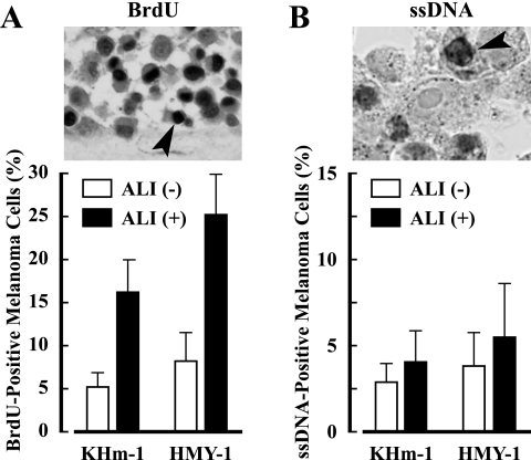
The air-liquid interface (ALI) is a common microenvironment of the skin, but it is unknown whether the ALI affects melanoma cell behaviors. Using a collagen gel invasion assay, immunohistochemistry, and Western blots, here we show that melanoma cell proliferation in cultures with an ALI is higher than melanoma cell proliferation in submerged cultures. Bromodeoxyuridine (BrdU) uptake, an indicator of cell proliferation, of melanoma cells at the ALI was about 3 times that of submerged cells, while ALI and submerged melanoma cells had similar levels of single-stranded DNA (a marker of apoptosis). The ALI enhanced the expression of Raf-1, MEK-1 and pERK-1/2 components of the mitogen-activated protein kinase (MAPK) cascade, in cells more than the submerged condition did. The increases in BrdU uptake and pERK-1/2 expression promoted by ALI was abolished by the MEK inhibitor, PD-98059. ALI-treated and submerged melanoma cells did not infiltrate into the collagen gel, and they showed no significant difference in the expression of the invasion- and motility-related molecules, matrix metalloproteinase-1 and -9, laminin 5, and filamin A. Our data indicate that the ALI, a skin microenvironment, accelerates the growth, but not the apoptosis or invasion, of melanoma cells through MAPK activation.

Keywords: air-liquid interface; collagen gel invasion assay; melanoma; mitogen-activated protein kinase; proliferation.


Figures

Fig. 1
Fig. 1
Collagen gel invasion assay system with (upper panel) or without ALI (lower panel). Upper panel, Melanoma cells are seeded on the acellular gel layer in inner dish (1). The inner dish (1) is put in outer dish (2) with culture medium. In this way, the cells on the gel layer are localized under air exposure-induced ALI. The cells are kept in moist and fed by culture medium that percolates by capillary action from the medium-containing outer dish. Lower panel indicates submerged culture without ALI (control). In these systems, the invasion of melanoma cell into collagen gel is analyzed by the method described in Materials and Methods.
Fig. 2
Fig. 2
Histology of two melanoma cell types of KHm-1 (A and B) and HMY-1 (C and D) with (B and D) or without ALI (A and C). ALI promotes the stratification of KHm-1 and HMY-1 melanoma cell types more greatly than submerged condition without ALI. In all conditions, the infiltration of these melanoma cell types into the gel was not observed. Brown pigments indicate melanin pigments.
Fig. 3
Fig. 3
Growth (A) and apoptosis (B) of melanoma cells with or without ALI, using immunohistochemistry of BrdU (growth marker) and ssDNA (apoptosis marker). BrdU (upper panel in A) and ssDNA (upper panel in B) are clearly detected in the nuclei of KHm-1 melanoma cells in black (arrowhead). The BrdU uptake of melanoma cells with ALI was about 3 times that of the cells without ALI (p<0.001), while melanoma cells with or without ALI showed no significant change in the apoptotic rate (p>0.05).
Fig. 4
Fig. 4
Expression of MAPK pathway (Raf-1, MEK-1 and ERK-1/2) of melanoma cells with or without ALI. In immunohistochemistry (A–F), KHm-1 melanoma cells with ALI (B, D and F) express Raf-1 (A and B), MEK-1 (C and D) and ERK-1/2 (E and F) more extensively than those without ALI (A, C and D). Western blotting (G) confirmed the immunohistochemical results. Analyses by densitometry (H and I) show that ALI significantly enhances Raf-1, MEK-1 and pERK-1/2 expression of KHm-1 (H) and HMY-1 (I) melanoma cells (all values, p<0.001). Total ERK-1/2 expression of melanoma cells with or without ALI had no significant change.
Fig. 5
Fig. 5
Expression of MMP-1, MMP-9, laminin 5 and filamin A of KHm-1 and HMY-1 melanoma cell types with or without ALI by Western blotting (A) and analyses by densitometry (B and C). MMP-1, MMP-9, laminin 5 and filamin A are weakly detected in KHm-1 and HMY-1 melanoma cells types with or without ALI. Analyses by densitometry show that ALI does not significantly affect these molecule expression of KHm-1 (B) and HMY-1 melanoma cell types (C) (all values, p>0.05).
Fig. 6
Fig. 6
Effects of the MEK inhibitor PD-98059 on the morphology, BrdU uptake and MAPK (ERK-1/2) expression of melanoma cells with ALI. ALI-treated KHm-1 melanoma cells without PD-98059 (A) undergo cellular stratification on the gel. In contrast, the melanoma cells with PD-98059 show no stratification and they become spindle-shaped (B). In ALI-treated KHm-1 and HMY-1 melanoma cell types, PD-98059 significantly inhibits both BrdU uptake (C, p<0.0001) and pERK-1/2 expression (D) of the cell types.

References

    1. Barnhill R. L., Gupta K. Unusual variants of malignant melanoma. Clin. Dermatol. 2009;27:564–587. - PubMed
    1. Bebok Z., Tousson A., Schwiebert L. M., Venglarik C. J. Improved oxygenation promotes CFTR maturation and trafficking in MDCK monolayers. Am. J. Physiol. Cell Physiol. 2001;280:C135–C145. - PubMed
    1. Bennett D. C. Genetics, development, and malignancy of melanocytes. Int. Rev. Cytol. 1993;146:191–260. - PubMed
    1. Bouffard D., Duncan L. M., Howard C. A., Mihm M. C., Jr., Byers H. R. Actin-binding protein expression in benign and malignant melanocytic proliferations. Hum. Pathol. 1994;25:709–714. - PubMed
    1. Brown D. Structure and function of membrane rafts. Int. J. Med. Microbiol. 2002;291:433–437. - PubMed