. 2010:2010:120328.
doi: 10.1155/2010/120328. Epub 2010 Mar 15.

ICAM-1 clustering on endothelial cells recruits VCAM-1

Affiliations

ICAM-1 clustering on endothelial cells recruits VCAM-1

Jaap D van Buul et al. J Biomed Biotechnol. 2010.

Abstract

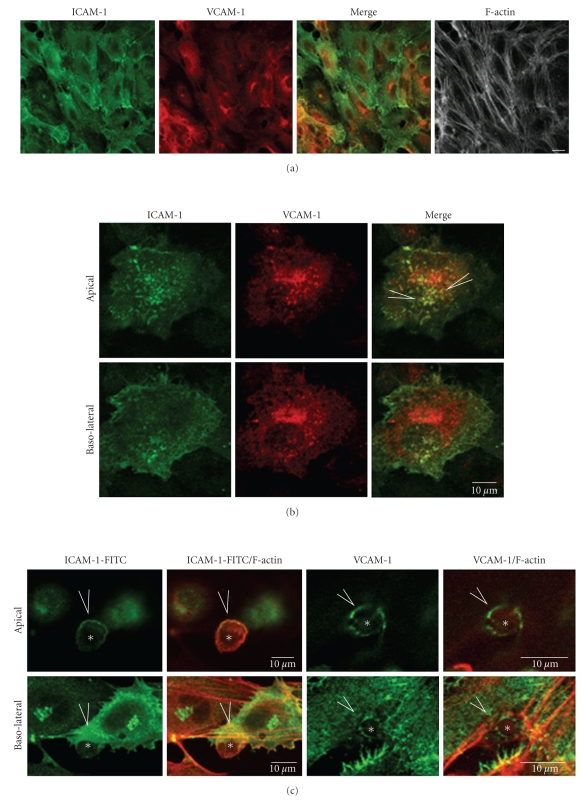
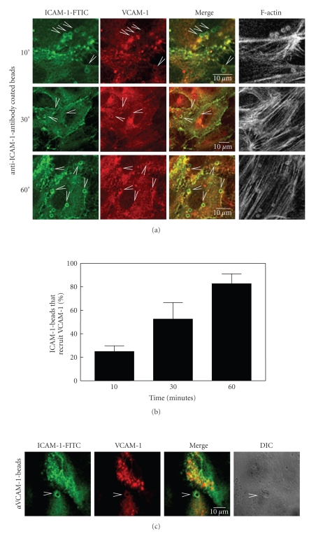
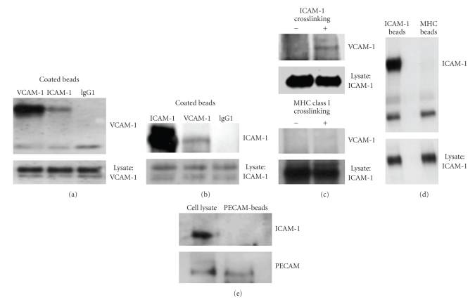
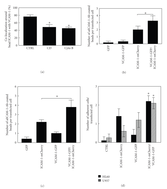
In the initial stages of transendothelial migration, leukocytes use the endothelial integrin ligands ICAM-1 and VCAM-1 for strong adhesion. Upon adhesion of the leukocyte to endothelial ICAM-1, ICAM-1 is clustered and recruited to the adhered leukocyte, promoting strong adhesion. In this study, we provide evidence for the colocalization of VCAM-1 at sites of ICAM-1 clustering. Anti-ICAM-1 antibody-coated beads were used to selectively cluster and recruit ICAM-1 on primary human endothelial cells. In time, co-localization of ICAM-1 and VCAM-1 around the adherent beads was observed. Biochemical pull-down assays showed that ICAM-1 clustering induced its association to VCAM-1, suggesting a physical link between these two adhesion molecules. The association was partly dependent on lipid rafts as well as on F-actin and promoted adhesion. These data show that VCAM-1 can be recruited, in an integrin-independent fashion, to clustered ICAM-1 which may serve to promote ICAM-1-mediated leukocyte adhesion.


Figures

Figure 1
Figure 1
ICAM-1 and VCAM-1 localization in TNFα-stimulated endothelial cells. (a) TNFα-treated HUVECs were fixed and stained as indicated. Bar, 20 μm. (b) Detailed analysis of ICAM-1 and VCAM-1 co-localization at the apical surface (arrowheads) in TNFα-treated endothelial cells that were fixed and stained as indicated. Bar, 10 μm. (c) Differentiated HL60 cells were allowed to adhere to TNFα-treated HUVECs for 30 minutes, after which cells were fixed and stained as indicated. Apical focal plane shows that ICAM-1 and VCAM-1 (arrowheads) are recruited around adhered HL60 cells (asterisks). Bar, 10 μm.
Figure 2
Figure 2
VCAM-1 is recruited to anti-ICAM-1-antibody coated beads. (a) ICAM-1 beads (diameter 3 μm) were allowed to adhere to TNFα-treated HUVECs. Subsequently, the samples were fixed at the indicated time points and stained as indicated. VCAM-1 is recruited to ICAM-1 beads (arrowheads) in time. Bar, 10 μm. (b) Quantification of VCAM-1 recruitment to ICAM-1-induced adhesion sites. The number of adhered ICAM-1-antibody beads that were positive for ICAM-1-FITC staining was set as 100% and percentage of beads that co-localized with VCAM-1 was counted. At 60 minutes adhesion, approximately 80% of ICAM-1 beads that recruited ICAM-1 also recruited VCAM-1. Data are mean ± SEM of three independent experiments. (c) VCAM-1 beads (diameter 3 μm) were allowed to adhere to TNFα-treated HUVECs and stained as indicated. ICAM-1 is recruited to VCAM-1 beads (arrowheads). Bar, 10 μm.
Figure 3
Figure 3
VCAM-1 associates with clustered ICAM-1. (a) Beads were coated with the indicated Abs as described in Section 2 and subsequently incubated with lysates from TNFα-treated HUVECs, followed by centrifugation, washing and analysis of bound proteins by SDS-PAGE and Western blotting. Blots were stained for VCAM-1 and show that VCAM-1 was precipitated with VCAM-1 beads and also with ICAM-1 beads. No VCAM-1 was detected in pull-downs using anti-IgG antibody coated beads. Lower panel shows expression of VCAM-1 in cell lysates as a loading control. Data are representative for three experiments. (b) The pull-down experiment was carried out as described under (a) and blots were stained for ICAM-1. ICAM-1 was precipitated with ICAM-1 beads, and a fraction of ICAM-1 associates with VCAM-1 beads, albeit less efficient than the reciprocal experiment shown in (a). No ICAM-1 was detected in pull-down experiments using anti-IgG coated beads. Lower panel shows expression of ICAM-1 as loading control in cell lysates. Data are representative for three experiments. (c) TNFα-stimulated HUVECs were incubated with ICAM-1 Ab in solution for 30 minutes and subsequently crosslinked with secondary antimouse Ab for an additional 30 minutes. Cells were lysed and ICAM-1 was immunoprecipitated and analyzed by Western blotting, which shows that ICAM-1-clustering induced association with VCAM-1. Lower panel shows ICAM-1 expression in cell lysates. Bottom panel shows that crosslinking of MHC class I protein did not induce any binding of VCAM-1. (d) ICAM-1 is precipitated by ICAM-1 beads, but not by anti-MHC class I antibody coated beads. Lower panel shows expression of ICAM-1 in cell lysates. (e) No ICAM-1 is precipitated by anti-PECAM-1 antibody coated beads (upper panel). Lower panel shows precipitation of PECAM-1 with the beads.
Figure 4
Figure 4
ICAM-1-induced VCAM-1 recruitment depends on lipid-rafts and actin polymerization and promotes ICAM-1-induced adhesion. (a) Anti-ICAM-1-antibody coated beads were allowed to adhere for 60 minutes to TNFα-stimulated HUVECs that were left untreated (CTRL) or were pretreated for 30 minutes with either cyclodextrin (CD; 3.8 μM) or cytochalasin B (CytoB; 1 μg/mL). Subsequently, cells were fixed and immunostained with ICAM-1-FITC and VCAM-1 Abs. VCAM-1 recruitment to ICAM-1 beads was quantified which showed that close to 80% of ICAM-1 beads which recruited ICAM-1 (set as 100%) also recruited VCAM-1. CD pretreatment reduced VCAM-1 recruitment with 40% and CytoB with 50%. Experiment was carried out three times. Data are mean ± SEM. *P < .05. (b) Anti-ICAM-1-antibody coated beads were allowed to adhere for 60 minutes to HeLa cells that were transfected as indicated. Subsequently, cells were fixed and analyzed by confocal microscopy for number of ICAM-1 beads that adhered to one transfected cell. 50 transfected cells per experiment were analyzed and the experiment is carried out in duplicate. The experiment was carried out three times. Data are mean ± SEM. *P < .05. (c) Anti-VCAM-1-antibody coated beads were allowed to adhere for 60 minutes to HeLa cells that were transfected as indicated. Subsequently, cells were fixed and analyzed by confocal microscopy for number of VCAM-1 beads that adhered to one transfected cell. 50 transfected cells per experiment were analyzed and the experiment is carried out in duplicate. The experiment was carried out three times. Data are mean ± SEM. *P < .05. (d) Nondifferentiated HL60 or U937 cells were allowed to adhere for 60 minutes to HeLa cells that were transfected as indicated. Subsequently, cells were fixed and analyzed by confocal microscopy for number of VCAM-1 beads that adhered to one transfected cell. 50 transfected cells per experiment were analyzed and the experiment is carried out in duplicate. The experiment was carried out two times. Data are mean ± SEM. *P < .05.

References

    1. Springer TA. Traffic signals for lymphocyte recirculation and leukocyte emigration: the multistep paradigm. Cell. 1994;76(2):301–314. - PubMed
    1. Kluger MS. Vascular endothelial cell adhesion and signaling during leukocyte recruitment. Advances in Dermatology. 2004;20:163–201. - PubMed
    1. van Buul JD, Mul FPJ, van der Schoot CE, Hordijk PL. ICAM-3 activation modulates cell-cell contacts of human bone marrow endothelial cells. Journal of Vascular Research. 2004;41(1):28–37. - PubMed
    1. Tilghman RW, Hoover RL. E-selectin and ICAM-1 are incorporated into detergent-insoluble membrane domains following clustering in endothelial cells. FEBS Letters. 2002;525(1–3):83–87. - PubMed
    1. Viola A, Gupta N. Tether and trap: regulation of membrane-raft dynamics by actin-binding proteins. Nature Reviews Immunology. 2007;7(11):889–896. - PubMed

Publication types

MeSH terms

Substances

LinkOut - more resources