. 2010 Mar 16;5(3):e9716.
doi: 10.1371/journal.pone.0009716.

In vivo accumulation of Helicobacter pylori products, NOD1, ubiquitinated proteins and proteasome in a novel cytoplasmic structure

Affiliations

In vivo accumulation of Helicobacter pylori products, NOD1, ubiquitinated proteins and proteasome in a novel cytoplasmic structure

Vittorio Necchi et al. PLoS One. .

Abstract

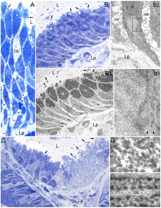
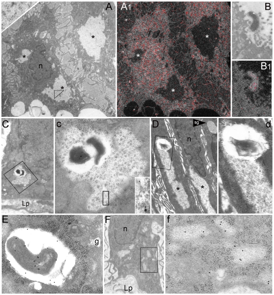
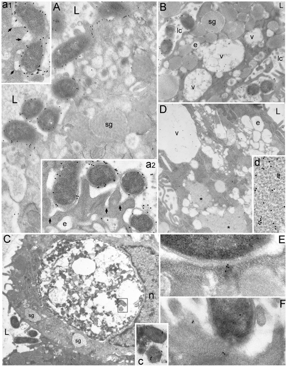
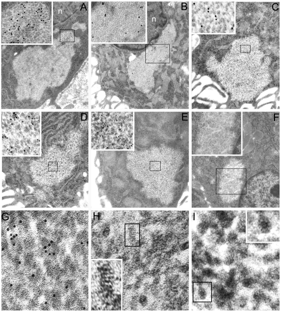
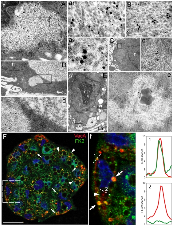
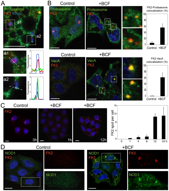
Cell internalization and intracellular fate of H. pylori products/virulence factors in vivo by human gastric epithelium, the main target of H. pylori-induced pathologies (i.e., peptic ulcer and cancer), are still largely unknown. Investigating gastric endoscopic biopsies from dyspeptic patients by means of ultrastructural immunocytochemistry, here we show that, in human superficial-foveolar epithelium and its metaplastic or dysplastic foci, H. pylori virulence factors accumulated in a discrete cytoplasmic structure characterized by 13-nm-thick cylindrical particles of regular punctate-linear substructure resembling the proteasome complex in size and structure. Inside this particle-rich cytoplasmic structure (PaCS) we observed colocalization of VacA, CagA, urease and outer membrane proteins with NOD1 receptor, ubiquitin-activating enzyme E1, polyubiquitinated proteins, proteasome components and potentially oncogenic proteins like SHP2 and ERKs in human gastric epithelium. By means of electron and confocal microscopy, we demonstrate that the in vivo findings were reproduced in vitro by incubating human epithelial cell lines with H. pylori products/virulence factors. PaCSs differed from VacA-induced vacuoles, phagosomes, aggresomes or related bodies. Our data suggest that PaCS is a novel, proteasome-enriched structure arising in ribosome-rich cytoplasm at sites of H. pylori products accumulation. As a site of selective concentration of bacterial virulence factors, the ubiquitin-proteasome system and interactive proteins, PaCS is likely to modulate immune-inflammatory and proliferative responses of the gastric epithelium of potential pathologic relevance.


Conflict of interest statement

Competing Interests: The authors have declared that no competing interests exist.

Figures

Figure 1
Figure 1. Identification of PaCS in H. pylori-colonized human gastric epithelium in vivo.
(A, 1,000x) and (B, 1,000x) H. pylori (arrows) colonized superficial gastric epithelium stained with toluidine blue shows metachromatic pink areas (arrowheads), mainly infranuclear, surrounded by blue stained (ribosome-rich) cytoplasm. Note in A a dendritic cell (DC; see [ref. 12]) approaching luminal bacteria. Aldehyde-osmium fixed, 0.5-µm-thick resin sections. (B1, 1,000x) TEM of the same epithelium in a consecutive section to B shows identity of the pink structures with PaCSs. A PaCS is enlarged in b2 (6,300x) and further in b3 (31,500x) to show the characteristic particles (left side in b3), to be compared with ribosomes of surrounding RER (right side in b3). Note in b3 direct contiguity of the PaCS with ribosomes and, in the lower right corner, with two RER cisternae (arrows), apparently without admixture of respective contents. (C, 500x) Toluidine blue stained resin section showing on the right a highly colonized, severely damaged epithelium with deeply irregular luminal border (due to cell bulging, desquamation and microerosion), vacuolation and loss of mucin granules in the apical-supranuclear cytoplasm, disappearance of ribosome-related basophilia in the basal cytoplasm and loss of cell polarity, to be compared with a relatively preserved epithelium in the lower left corner and a moderately damaged epithelium upper left. Note several metachromatic areas in lower left (arrowheads), a single fainty stained area upper left and no metachromatic areas in the severely damaged epithelium on the right. (D, 120,000x) High-resolution TEM of barrel-like, randomly oriented PaCS particles, 13 nm thick and 15 to 43 nm long, to show their regular punctate substructure. Compare with more dense, frequently angular ribosomes aligned along RER cisternae in E (120,000x). cs, cisterna; ics, intercellular space; L, gastric lumen; Lp, lamina propria; n, nucleous; sg, secretory granules.
Figure 2
Figure 2. Characterization of PaCS in H. pylori-colonized human gastric epithelium in vivo.
(A) and (A1) (both 4,000x) In two foveolar cells (A) three PaCSs (asterisks), one of which enlarged in the inset (upper left corner, 50,000x) to show their particles, lack phosphorus signal (red colour) when viewed under ESI analysis (A1). Note the intense signal of nucleoproteins in the nucleous, cytoplasmic ribosomes surrounding PaCSs and, in B and B1 (both 8,000x), a luminal H. pylori adhering to epithelial cell microvilli. (C, 6,300x) and (D, 6,300x) H. pylori bodies inside PaCSs, two of which fairly-well preserved (C; enlarged in c, 16,000x), the other (D; enlarged in d, 16,000x) heavily degenerated. Note a peribacterial clear space, the immunogold reactivity of both bacteria and PaCS for H. pylori OMPs (5 nm gold) and VacA (10 nm gold) in c and its inset (44,000x), while the PaCS only, but not bacterial remnants, reacts for human 20S-β5i subunit of the immunoproteasome (d). (E, 25,000x) OMP-reactive intracellular H. pylori enclosed in a supranuclear vacuole of a foveolar cell. (F, 6,300x) Several small, thin PaCSs are sparse inside the RER at the base of an epithelial cell, enlarged in f (32,000x) to show their particles clearly less dense than ribosomes, as well as their selective immunogold reactivity for CagA. g, Golgi area; Lp, lamina propria; n, nucleous; sg, secretory granule.
Figure 3
Figure 3. CagA, urease, VacA, and polyubiquitinated proteins accumulate in PaCS.
Foveolar-superficial cells with luminal H. pylori localization, in the apparent absence of intracellular bacteria (A, 8,000x), show several PaCSs with selective immunoreactivity for: CagA (enlarged in a1, 12,000x, and in a2, 84,000x); urease (B, 8,000x), with PaCSs on both sides of the nucleous, one of which enlarged in b (84,000x); VacA (C, 4,500x; one enlarged in c, 60,000x); and polyubiquitinated proteins (D, 9,500x; enlarged in d1, 12,000x, and further in d2, 84,000x). Note in A and D remnants of cytoplasm inside PaCSs, suggesting their origin from enlargement and fusion of smaller structures like those in Figure 2F. L, gastric lumen; Lp, lamina propria; n, nucleous.
Figure 4
Figure 4. PaCSs differ from VacA-containing vacuoles and phagosomes.
(A, 12,000x, 10 nm gold), (a1, 20,000x), and (a2, 20,000x) H. pylori organisms closely adhering to the luminal surface of colonized cells in superficial gastric epithelium. Note VacA immunoreactivity in bacterial periplasm, cellular early carriers under formation immediately below bacterial adhesion (arrows) and some apical endocytic-endosomal vesicles. (B, 7,000x) Endoluminal bleb of a colonized superficial cell showing immunoreactive VacA in adhering bacteria and some endosomal vesicles fusing each other to form VacA-storing larger vacuoles with inner remnants of original endosomal membranes. (C, 10,000x) Colonized cell showing a large supranuclear phagosome with abundant cytoplasmic remnants and debris and VacA immunoreactivity, enlarged in c (40,000x). (D, 7,000x) HeLa cells incubated for 24 h with H. pylori BCF. Note inside the cell VacA immunoreactivity of some endosomal vesicles, a large vacuole as well as two PaCSs (asterisks) with distinctive particles (d, 60,000x). (E) and (F) (both 55,000x) Small CagA deposits in the subluminal cytoplasm just below two adhering H. pylori. L, gastric lumen; foveolar cell type; e, endosomal vesicle; lc, luminal cleft; n, nucleous; sg, secretory granules; v, vacuole.
Figure 5
Figure 5. Proteasome is the particle component of PaCS which also contains NOD1.
Selective PaCS reactivity for NOD1 (A, 12,500x; boxed part enlarged in its inset, 60,000x); ubiquitin-activating enzyme E1 (B, 10,000x; enlarged in its inset, 32,000x); 20S proteasome (C, 12,500x; enlarged in its inset, 60,000x); 20S-β5i subunit of immunoproteasome (D, 12,500x; enlarged in its inset, 60,000x); and 19S proteasome (E, 12,500x; enlarged in its inset, 60,000x). Note in F (10,000x) and its inset (32,000x) lack of immunogold particles in a PaCS and surrounding cytoplasm of a control section from the same resin block (as in A, B, and C) incubated with gold-labelled non-immune globulins. In G (125,000x), H (340,000x) and I (340,000x) high-resolution TEM of the 13-nm-thick PaCS particles, selectively immunoreactive for 19S proteasome (G), shows their inner punctate substructure with minute electrondense spots aligned perpendicularly to particle long axis. On a side view like that further enlarged in the inset of H (600,000x), some particles closely resemble the four parallel rings of a proteasome 20S core capped at both estremities, while on a top view like that in the inset of I (600,000x) they may reproduce the proteasome seven-fold star-like symmetry. n, nucleous.
Figure 6
Figure 6. Colocalization in PaCS of bacterial products, UPS and oncogenic/signaling molecules in vivo and in vitro.
(A, 20,000x; enlarged in a1, 95,000x, and a2, 120,000x) Colocalization in PaCSs of CagA (15 nm gold particles) with polyubiquitinated proteins (10 nm gold particles) and 20S-β5i subunit of immunoproteasome (5 nm gold particles). Note in a1 and a2 close reciprocal contacts of some 20-nm gold particles with 10- or 15-nm ones; also note in a2 the linear-punctate substructure of PaCS particles. (B, 80,000x) In another section through the same PaCS as of A, colocalization of CagA (15 nm gold) and SHP2 protein (10 nm gold) is obtained. (C, 5,000x; enlarged in c, 35,000x) Another PaCS showing selective ERK reactivity. (D, 10,000x; enlarged in d, 45,000x) In addition to a typical particulate PaCS (left part of d) immunoreactive for FK1 antibodies (recognizing polyubiquitinated proteins), another, FK1-unreactive non-particulate cytoplasmic structure (upper right of D and d) with a filamentous-honeycomb meshwork. (E, 6,300x) Dysplastic cell with prominent nucleolus showing several PaCSs (one labelled with asterisk) in its basal cytoplasm. A PaCS containing bacterial remnants is enlarged in e (20,000x) to show H. pylori (OMPs) immunoreactivity of both bacterial body and PaCS itself. n, nucleous; nl, nucleolus. (F) Confocal microscopy of HeLa cells (nuclei in blue) incubated for 24 h with H. pylori BCF shows the presence in the cytoplasm of yellow spots (arrows; see also enlargement in f) representing the colocalization of ubiquitinated proteins (FK2; green) with VacA (red). Note that vacuole-associated VacA (arrowheads) does not colocalize with FK2. Colocalization charts (graphs 1 and 2) show the intensity profile for each fluorescence taken along the dotted lines. Pictures are from one single confocal section. Bar: 25 µm.
Figure 7
Figure 7. H. pylori products/virulence factors induce PaCS formation in vitro.
(A) Confocal microscopy of HeLa cells incubated for 24 h with Cy5-labeled purified VacA for the simultaneous detection of proteasome (green), ubiquitinated proteins (FK2; red), and VacA (blue). The enlargements show in a1 colocalization (purple) between ubiquitinated proteins and VacA, and in a2 colocalization (light blue/white) of proteasome, ubiquitinated proteins, and VacA. The graphs on the right represent the respective colocalization charts showing the intensity profile for each of the three fluorescences taken along the dotted lines. Pictures are from one single confocal section. Bar: 10 µm. (B) Confocal microscopy of HeLa cells (nuclei in blue) incubated for 24 h without (control) or with H. pylori BCF shows the presence in the cytoplasm of BCF-treated cells of spots with the yellow component representing the colocalization of ubiquitinated proteins (FK2; red) with proteasome (green) (top line) or of ubiquitinated proteins (red) with VacA (green) (bottom line), respectively. These cytoplasmic colocalizations in either control or treated cells are quantitatively analyzed in the histograms (right) showing the percentage of FK2-positive spots colocalizing with proteasome or VacA, respectively. Pictures are from one single confocal section. Bar: 10 µm. (C) Time-course of HeLa cells (nuclei in blue) incubated with H. pylori BCF shows a time-dependent increase in the cytoplasmic spots of ubiquitinated proteins (FK2; red). The histogram (right) shows the number (means ± SEM) of FK2-positive cytoplasmic spots per cell. Bar: 10 µm. (D) Confocal microscopy of HeLa cells (nuclei in blue) incubated for 24 h without (control) or with H. pylori BCF shows the presence in the cytoplasm of BCF-treated cells of yellow spots representing the colocalization of ubiquitinated proteins (FK2; red) with NOD1 (green). Each individual labeling is shown in the enlarged areas of the boxed regions. Pictures are from one single confocal section. Bar: 10 µm.

Similar articles

Cited by

References

    1. Chan WY, Hui PK, Leung KM, Thomas TM. Modes of Helicobacter colonization and gastric epithelial damage. Histopathology. 1992;21:521–528. - PubMed
    1. Necchi V, Candusso ME, Tava F, Luinetti O, Ventura U, et al. Intracellular, intercellular, and stromal invasion of gastric mucosa, preneoplastic lesions, and cancer by Helicobacter pylori. Gastroenterology. 2007;132:1009–1023. - PubMed
    1. Amieva MR, Salama NR, Tompkins LS, Falkow S. Helicobacter pylori enter and survive within multivesicular vacuoles of epithelial cells. Cell Microbiol. 2002;4:677–690. - PubMed
    1. Odenbreit S, Püls J, Sedlmaier B, Gerland E, Fischer W, et al. Translocation of Helicobacter pylori CagA into gastric epithelial cells by type IV secretion. Science. 2000;287:1497–1500. - PubMed
    1. Stein M, Rappuoli R, Covacci A. Tyrosine phosphorylation on the Helicobacter pylori CagA antigen after cag-driven host cell translocation. Proc Natl Acad Sci USA. 2000;97:1263–1268. - PMC - PubMed

Publication types

Substances