. 2010 Mar 12;6(3):e1000802.
doi: 10.1371/journal.ppat.1000802.

Membrane damage elicits an immunomodulatory program in Staphylococcus aureus

Affiliations

Membrane damage elicits an immunomodulatory program in Staphylococcus aureus

Ahmed S Attia et al. PLoS Pathog. .

Abstract

The Staphylococcus aureus HrtAB system is a hemin-regulated ABC transporter composed of an ATPase (HrtA) and a permease (HrtB) that protect S. aureus against hemin toxicity. S. aureus strains lacking hrtA exhibit liver-specific hyper-virulence and upon hemin exposure over-express and secrete immunomodulatory factors that interfere with neutrophil recruitment to the site of infection. It has been proposed that heme accumulation in strains lacking hrtAB is the signal which triggers S. aureus to elaborate this anti-neutrophil response. However, we report here that S. aureus strains expressing catalytically inactive HrtA do not elaborate the same secreted protein profile. This result indicates that the physical absence of HrtA is responsible for the increased expression of immunomodulatory factors, whereas deficiencies in the ATPase activity of HrtA do not contribute to this process. Furthermore, HrtB expression in strains lacking hrtA decreases membrane integrity consistent with dysregulated permease function. Based on these findings, we propose a model whereby hemin-mediated over-expression of HrtB in the absence of HrtA damages the staphylococcal membrane through pore formation. In turn, S. aureus senses this membrane damage, triggering the increased expression of immunomodulatory factors. In support of this model, wildtype S. aureus treated with anti-staphylococcal channel-forming peptides produce a secreted protein profile that mimics the effect of treating DeltahrtA with hemin. These results suggest that S. aureus senses membrane damage and elaborates a gene expression program that protects the organism from the innate immune response of the host.


Conflict of interest statement

The authors have declared that no competing interests exist.

Figures
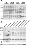
Figure 1
Figure 1. Up-regulation of immunomodulatory proteins is caused by loss of expression of HrtA.
(A) Bacterial strains were grown in (0, 0.5 and 2 µM) hemin in RPMI/CAS for 18 hours and proteins in culture supernatants were precipitated using 10% TCA, separated using 15% SDS-PAGE, and stained with Coomassie blue. (B) Exoprotein profiles of wildtype transformed with control plasmid pOS1 and ΔhrtA transformed with control plasmid or plasmids expressing WT HrtA, catalytically inactive HrtA variants (K45A, G145A, G145T, and E167Q) or partially catalytically active HrtA (R76A) . All strains were grown in the presence of 1 µM hemin. Proteins up-regulated under the indicated condition that have been identified previously using mass spectrometry are marked with a #. The predicted identities of the proteins in these bands are as follows; #1 (Exotoxin 8, SACOL0472), #2 (Exotoxin, SACOL0478), #3 (Exotoxin 3 and 5, SACOL0468/0473), #4 (Efb, SACOL1168), and #5 (FLIPr, SACOL1166). Positions of protein molecular mass markers in kilodaltons (kDa) are indicated on the left side of panels A and B. (C) Immunoblot analysis of protoplast lysates of strains analyzed in (B). Proteins were resolved using 15% SDS-PAGE, transferred to nitrocellulose membrane, probed with 9E10 anti-C-Myc monoclonal primary and AlexaFluor-680-conjugated anti-mouse secondary antibodies. Membranes were then dried and scanned using an Odyssey Infrared Imaging System.
Figure 2
Figure 2. Loss of HrtB alone does not affect protein secretion or virulence in mice.
(A) Diagrammatic representation of the hrtAB locus; the relative positions of the primers used for RT-PCR are indicated by black arrows. (B) Agarose gels showing the results of an RT-PCR experiment involving the hrtAB locus and the ribosomal 16S RNA as the loading control. Bacteria were grown ±1 µM hemin and RT-PCRs were done using DNA as the template (DNA), RNA with no reverse transcriptase (No RT) and RNA with reverse transcriptase (+RT). (C) Exoprotein profile of ΔhrtA + 1 µM hemin and ΔhrtB ±1 µM hemin. The # indicates the positions of proteins up-regulated under the indicated condition and the predicted identity of these proteins is as described in Figure 1. Positions of protein molecular mass markers in kilodaltons (kDa) are shown on the left side of the gel. (D) Bacterial multiplication in infected BALB/c mouse livers as measured by tissue homogenization, dilution, and colony formation on agar media 96 hours post infection. The horizontal bars represent the mean of log10CFU, and the boxes cover the range of log10CFU obtained in each group. * indicates statistically significant differences from ΔhrtA as determined by Student's t test with the indicated p values. NS indicates a non-statistically significant difference. N indicates the number of mice included in each group.
Figure 3
Figure 3. Over-expression of HrtB without hemin exposure mimics the effect of exposing ΔhrtA to hemin.
(A and B) FACS analysis of wildtype (A) and ΔhrtA (B) cells incubated ±1 µM hemin and stained with propidium iodide (PI). The experiment was performed at least three times and a representative result is presented. (C) Viable counts of serial dilutions of cell suspensions analyzed in (A and B). (D) Immunoblot analysis of protoplast lysates of strains transformed with either control plasmid pOS1-plgt (−) or plasmid encoding constitutively expressed Myc-tagged HrtB phrtB-myc (+). Immunoblots were analyzed as described in Figure 1C. (E) Exoprotein profile of the indicated strains were prepared and analyzed as in Figure 1. The # indicates the positions of proteins up-regulated under the indicated condition and the predicted identity of these proteins is as described in Figure 1. (F) Immunoblot analysis of cell fractions of WT/phrtB-myc. Equivalent amounts of proteins were loaded from each fraction and the immunoblot was analyzed as described in Fig. 1C The reactive band in the cell wall fraction is the S. aureus protein A (Spa), which reacts non-specifically with anti-c-Myc antibody. Positions of protein molecular mass markers in kilodaltons (kDa) are indicated on the left side of panels D and E.
Figure 4
Figure 4. Membrane pore formation but not generalized membrane damage mimics the effect of hemin on ΔhrtA.
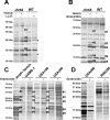
(A) Exoprotein profile of ΔhrtA treated ±1 µM hemin and WT ±40 µg/ml of the antimicrobial peptide LL37. (B) Exoprotein profile of ΔhrtA treated ±1 µM hemin and WT ±16 µg/ml of the antimicrobial peptide gramicidin. (C) Exoprotein profile of ΔhrtA treated with 1 µM hemin and different staphylococcal strains ±32 µg/ml gramicidin. (D) Exoprotein profile of wildtype strains USA500 and RN6390 ±32 µg/ml gramicidin. Samples were prepared and analyzed as in Figure 1. The # indicates the positions of proteins up-regulated under the indicated condition and the predicted identity of these proteins is as described in Figure 1. The * indicates the positions of proteins down-regulated under the indicated condition. Positions of protein molecular mass markers in kilodaltons (kDa) are indicated on the left side of each panel.
Figure 5
Figure 5. Treatment of S. aureus wildtype with gramicidin and ΔhrtA with hemin elicit similar secretomes.
(A) Average number of spectra per protein of secreted proteins that were significantly up-regulated in ΔhrtA + 1 µM hemin and WT + 32 µg/ml gramicidin. (B) Average number of spectra per protein of secreted proteins that were significantly down-regulated under both conditions. The data presented are means of three independent samples and the error bars represent the standard deviation. The differences between ΔhrtA + hemin (black bars) and ΔhrtA - hemin (white bars) and WT + gramicidin (diagonally streaked bars) and WT - gramicidin (grey bars) are statistically significant in all the presented proteins with p values less than 0.05 as indicated by Student's t test. * T stands for truncated. (C-F) Results of quantitative real-time RT-PCR using RNA samples isolated from samples of ΔhrtA ± hemin and WT ± gramicidin. The levels of the transcripts were normalized using the levels of the ribosomal RNA 16S and the levels of the normalized transcripts from samples without treatment (either hemin or gramicidin) were used as calibrators. The data presented are the means of two independent experiments with each done in triplicate and the error bars represent the standard deviation. The differences between ΔhrtA + hemin (black bars) and ΔhrtA - hemin (white bars) and WT + gramicidin (diagonally streaked bars) and WT - gramicidin (grey bars) are statistically significant in all the presented transcripts with p values less than 0.05 as indicated by Student's t test except the one data point marked with NS.
Figure 6
Figure 6. Ssls1-11 and Ssp are responsible for the liver-specific hyper-virulence of hrtA mutants.
(A) Growth curve analysis of wildtype, thrtA, thrtAΔssls, thrtAΔssp, and thrtAΔsslsΔssp. Overnight cultures were diluted 1:20 in TSB and the absorbance at 600 nm was recorded at the indicated time points. A representative growth curve is presented. (B) Bacterial multiplication in infected BALB/c mice livers as measured by tissue homogenization, dilution, and colony formation on agar media 96 hours post infection. The horizontal bars represent the mean of log10CFU, and the boxes cover the range of log10CFU obtained in each group. * indicates statistically significant differences from thrtA as determined by Student's t test with the indicated p values. NS indicates a non-statistically significant difference. N indicates the number of mice included in each group.
Figure 7
Figure 7. Membrane damage triggers immunomodulatory proteins secretion.
(A) When ΔhrtA is exposed to hemin (1) the HssS/R system is activated (2) leading to over-expression of the hrtB gene (3) causing membrane damage by the over-expressed HrtB proteins acting as unregulated pores (4). This in turn activates an internal signal (5) that turns on the expression (6) and secretion (7) of the immunomodulatory proteins. (B) When WT S. aureus is attacked by antimicrobial peptides (1) that form pores in the membrane (2), this in turn activates an internal signal (3) that turns on the expression (4) and secretion (5) of immunomodulatory proteins.

References

    1. Wertheim HF, Vos MC, Ott A, van Belkum A, Voss A, et al. Risk and outcome of nosocomial Staphylococcus aureus bacteraemia in nasal carriers versus non-carriers. Lancet. 2004;364:703–705. - PubMed
    1. Diekema DJ, Pfaller MA, Schmitz FJ, Smayevsky J, Bell J, et al. Survey of infections due to Staphylococcus species: frequency of occurrence and antimicrobial susceptibility of isolates collected in the United States, Canada, Latin America, Europe, and the Western Pacific region for the SENTRY Antimicrobial Surveillance Program, 1997-1999. Clin Infect Dis. 2001;32(Suppl 2):S114–132. - PubMed
    1. Klevens RM, Morrison MA, Nadle J, Petit S, Gershman K, et al. Invasive methicillin-resistant Staphylococcus aureus infections in the United States. JAMA. 2007;298:1763–1771. - PubMed
    1. Lowy FD. Staphylococcus aureus infections. N Engl J Med. 1998;339:520–532. - PubMed
    1. DeLeo FR, Diep BA, Otto M. Host defense and pathogenesis in Staphylococcus aureus infections. Infect Dis Clin North Am. 2009;23:17–34. - PMC - PubMed

Publication types

MeSH terms