Case Reports
. 2010 Mar 12;6(3):e1000874.
doi: 10.1371/journal.pgen.1000874.

Fatal cardiac arrhythmia and long-QT syndrome in a new form of congenital generalized lipodystrophy with muscle rippling (CGL4) due to PTRF-CAVIN mutations

Affiliations
Case Reports

Fatal cardiac arrhythmia and long-QT syndrome in a new form of congenital generalized lipodystrophy with muscle rippling (CGL4) due to PTRF-CAVIN mutations

Anna Rajab et al. PLoS Genet. .

Abstract

We investigated eight families with a novel subtype of congenital generalized lipodystrophy (CGL4) of whom five members had died from sudden cardiac death during their teenage years. ECG studies revealed features of long-QT syndrome, bradycardia, as well as supraventricular and ventricular tachycardias. Further symptoms comprised myopathy with muscle rippling, skeletal as well as smooth-muscle hypertrophy, leading to impaired gastrointestinal motility and hypertrophic pyloric stenosis in some children. Additionally, we found impaired bone formation with osteopenia, osteoporosis, and atlanto-axial instability. Homozygosity mapping located the gene within 2 Mbp on chromosome 17. Prioritization of 74 candidate genes with GeneDistiller for high expression in muscle and adipocytes suggested PTRF-CAVIN (Polymerase I and transcript release factor/Cavin) as the most probable candidate leading to the detection of homozygous mutations (c.160delG, c.362dupT). PTRF-CAVIN is essential for caveolae biogenesis. These cholesterol-rich plasmalemmal vesicles are involved in signal-transduction and vesicular trafficking and reside primarily on adipocytes, myocytes, and osteoblasts. Absence of PTRF-CAVIN did not influence abundance of its binding partner caveolin-1 and caveolin-3. In patient fibroblasts, however, caveolin-1 failed to localize toward the cell surface and electron microscopy revealed reduction of caveolae to less than 3%. Transfection of full-length PTRF-CAVIN reestablished the presence of caveolae. The loss of caveolae was confirmed by Atomic Force Microscopy (AFM) in combination with fluorescent imaging. PTRF-CAVIN deficiency thus presents the phenotypic spectrum caused by a quintessential lack of functional caveolae.


Conflict of interest statement

The authors have declared that no competing interests exist.

Figures

Figure 1
Figure 1. Phenotype of patient FI:201 from Oman.
(A) Patient at the age of 4 years with macroglossia, grossly reduced subcutaneous fat tissue and a protruding abdominal wall. (B) ECG of the patient with a QTc (Bazett) of 480 ms. (C) Sections of 24h ECG Holter-monitoring show a complex cardiac arrhythmia with intermittent sinus bradycardia, supraventricular (SVT) and ventricular tachycardia (VT). BPM, beats per minute (D) Percussion-induced, local prolonged contractions (“mounding”) at the quadriceps muscle persisting for 2–3 seconds. (E) Pedigree of the consanguineous family and genotypes of the family members. (F) X-ray radiograph of the knees at 9 months of age, showing broadening of the distal metaphyses (arrowheads) and osteopenia. (G) X-ray radiograph of the left hand at the age of 13.5 years showing osteoporosis and osteopenia. The metacarpo-phalangeal joint of the thumb shows arthritic changes and a partial dislocation with ulnar deviation (arrowhead).
Figure 2
Figure 2. Phenotype of patient FII:201 from the UK.
(A) Image of the 12-year-old patient with generalized lack of subcutaneous fat and prominent muscle hypertrophy especially of the thigh (see also Figure S1), masticatory and paraspinal muscles. The patient presented with spinal rigidity and lumbar hyperlordosis. The veins generally appeared thickened and prominent (phlebomegaly). Atlanto-axial instability of the patient during flexion (B), and extension (C) of the cervical spine. In flexion, the gap between the anterior arc of the atlas (dotted triangle) and the odontoid process of the axis (closed triangle) opens up 7 mm. The posterior arch of the axis appears dysplastic. During flexion the posterior margins of the cervical vertebrae are misaligned (dotted line). There is marked loss of bone mineral density and increase of translucency of the cervical vertebrae due to osteoporosis. (D) Pedigree of the consanguineous family and genotypes of the family members. (E) ECG of the patient with a QTc (Bazett) of 501 ms.
Figure 3
Figure 3. Abnormal fat distribution in patients with PTRF-CAVIN mutations.
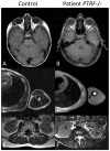
T1-weighted MR-images of patient FI:201 (B,D) and of patient FII:201 (F). On the left, the corresponding images of normal controls (A,C,E) are shown for comparison. T1-weighted images depict fat with high signal intensity thus giving an overview of the fat distribution. Subcutaneous fat is nearly completely lost over the peripheries, thoracic and abdominal walls and on both temporal regions. There is relative preservation of fat within the orbits. Paraspinal and perirenal fat is also reduced in bulk but relatively preserved. Fat in the bone marrow of the ribs and the humerus seems to be normal.
Figure 4
Figure 4. Family pedigrees and molecular genetic characterization of the PTRF-CAVIN mutations.
(A) Pedigrees of all investigated family members. With the exception of Family II, who came from the United Kingdom, all other families originated from Oman. The red arrowheads indicate the patients included for homozygosity mapping. The genotype of each individual is marked below the symbol. (B–D) Molecular findings in Family I in whom we found a deletion of a guanine residue at nt160 (hatched box) leading to a shift in the open reading frame. (C) Verification of the mutation by restriction enzyme analysis. The mutation creates a MwoI restriction site cutting the 137 bp band into 67+70 bp if the mutation is present. (D) Western blot of cultured fibroblasts from patient FI:201. PTRF-CAVIN immunoreactivity is completely absent, whereas caveolin-1 immunoreactivity is normal. The bottom panel shows the β-tubulin staining as loading control. (E–G) Molecular findings of Family II, in whom we discovered a duplication of a thymine residue at position 362 (hatched box) with subsequent frameshift. (F) Verification of the mutation by restriction analysis. In the presence of the mutation AclI cleaves a 539 bp band into 415+124 bp. (G) Western blot of muscle tissue from patient FII:201. Again, PTRF-CAVIN is completely absent from muscle, whereas there is no difference in caveolin-3 immunoreactivity. The bottom panel shows the myosin band as the loading control.
Figure 5
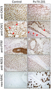
Figure 5. Immunohistochemistry of a muscle biopsy specimen from patient FII:201.
In the patient caveolin-3 expression in skeletal muscle fibers was reduced and irregular; caveolin-1 staining of intramuscular fat cells (red arrowheads) was completely absent. In the control, strong PTRF-staining can be seen in the walls of the small arterioles, representing the smooth muscle layer. It is virtually absent in the patient, nota bene: The nuclear staining of the anti-PTRF-antibody is unspecific. In the patient the intensity of the subsarcolemmal anti-NOS1 staining seems to be stronger and less patchy than in the control muscle. Overall the muscle of the patient shows myopathic changes, mild variation in fiber sizes without necrosis, inflammation or fibrosis and increased regeneration. These regenerating fibers (ca. 25%) are marked through positive staining for neo-MHC, an isofom of the myosin heavy chain protein that is characteristically expressed in neonatal muscle and in regenerating fibers.
Figure 6
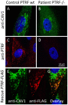
Figure 6. Cell-biological consequences of PTRF-CAVIN depletion.
(A) Confocal microscopic image of the punctate distribution of caveolin-1 which labels the caveolae on the surface of a fibroblast. (B) Severe reduction of the punctate caveolin-1 distribution in the absence of PTRF-CAVIN and its unstructured distribution within the cytoplasm. (C) Normal punctate distribution of PTRF-CAVIN on the fibroblast surface. (D) Absence of all PTRF-CAVIN immunoreactivity on a patient fibroblast. (E–G) Each panel depicts two patient fibroblasts, one untransfected (right) and one transfected with PTRF-FLAG construct (left). In the untransfected cell caveolin-1 is only found in the Golgi-apparatus. Reexpression of PTRF-CAVIN in the left cell redirects the caveolin-1 staining to the caveolae in the cell periphery where the two proteins co-localize (yellow dots in panel G).
Figure 7
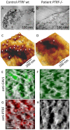
Figure 7. Ultrastructural analysis of caveolae on the fibroblast surface.
(A,B) Transmission electron micrographs (x 37.000) of ultrathin sections from near the cell surface of a fibroblast monolayer. The cell surface is labeled with ruthenium red. In the control individual (A) numerous caveolae and indentations of the cell membrane were seen close to the cell surface. (B) In the fibroblasts of patient FI:201 only rarely coated invaginations could be found. (C,D) High resolution Atomic Force Microscopic (AFM) scans of an area of 1×1 µm on the surface of (C) control and (D) patient fibroblasts. The caveolae (white arrowheads) have a size between 50–100 nm and are predominantly located at the margins of major membrane folds. The patient fibroblasts show a smooth surface with only occasional caveolae. (E–H) Overlay of AFM-images with the respective fluorescent immunostainings from the same surface plane. On the AFM-images one can localize the caveolae by the punctate caveolin-1 staining within larger indentations of the cell membrane which disappear in the absence of PTRF-CAVIN.

Similar articles

Cited by

References

    1. Garg A. Acquired and inherited lipodystrophies. N Engl J Med. 2004;350:1220–1234. - PubMed
    1. Garg A, Agarwal AK. Lipodystrophies: Disorders of adipose tissue biology. Biochim Biophys Acta. 2009;1791:507–513. - PMC - PubMed
    1. Magre J, Delepine M, Khallouf E, Gedde-Dahl T, Jr, Van Maldergem L, et al. Identification of the gene altered in Berardinelli-Seip congenital lipodystrophy on chromosome 11q13. Nat Genet. 2001;28:365–370. - PubMed
    1. Agarwal AK, Arioglu E, De Almeida S, Akkoc N, Taylor SI, et al. AGPAT2 is mutated in congenital generalized lipodystrophy linked to chromosome 9q34. Nat Genet. 2002;31:21–23. - PubMed
    1. Kim CA, Delepine M, Boutet E, El Mourabit H, Le Lay S, et al. Association of a homozygous nonsense caveolin-1 mutation with Berardinelli-Seip congenital lipodystrophy. J Clin Endocrinol Metab. 2008;93:1129–1134. - PubMed

Publication types

MeSH terms