. 2010 Mar 12;6(3):e1000879.
doi: 10.1371/journal.pgen.1000879.

MiR-218 inhibits invasion and metastasis of gastric cancer by targeting the Robo1 receptor

Affiliations

MiR-218 inhibits invasion and metastasis of gastric cancer by targeting the Robo1 receptor

Jun Tie et al. PLoS Genet. .

Abstract

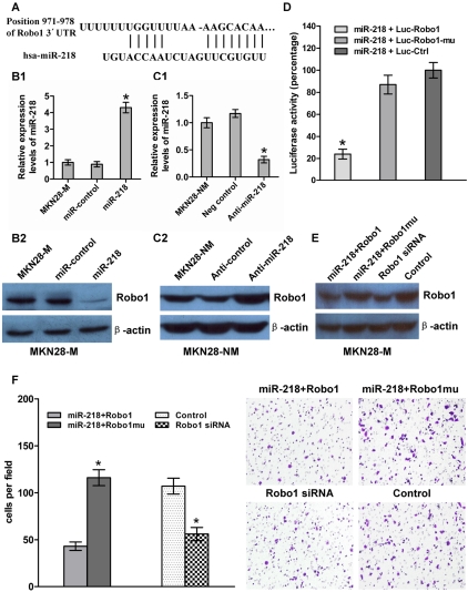
MicroRNAs play key roles in tumor metastasis. Here, we describe the regulation and function of miR-218 in gastric cancer (GC) metastasis. miR-218 expression is decreased along with the expression of one of its host genes, Slit3 in metastatic GC. However, Robo1, one of several Slit receptors, is negatively regulated by miR-218, thus establishing a negative feedback loop. Decreased miR-218 levels eliminate Robo1 repression, which activates the Slit-Robo1 pathway through the interaction between Robo1 and Slit2, thus triggering tumor metastasis. The restoration of miR-218 suppresses Robo1 expression and inhibits tumor cell invasion and metastasis in vitro and in vivo. Taken together, our results describe a Slit-miR-218-Robo1 regulatory circuit whose disruption may contribute to GC metastasis. Targeting miR-218 may provide a strategy for blocking tumor metastasis.


Conflict of interest statement

The authors have declared that no competing interests exist.

Figures

Figure 1
Figure 1. The metastatic characteristics of each cell subline.
(A) General scheme of the establishment of invasive and non-invasive cell sublines derived from human GC cell lines. (B,C) In vitro migration and invasion activity of each cell subline. Migration and invasion activities were measured in vitro with transwell chambers, as described in Materials and Methods. Photos are representative fields of invasive cells on the membrane. Magnification, 100x. Bar graphs represent the average number of cells on the underside of the membrane ± SE. ** P<0.01 as compared with non-invasive cells, analyzed by t-test. (D and E) Metastasis potential of each cell subline in vivo (n = 10). (D) The incidence of metastasis in mice that received intravenous tail injections of each selected cell subline. (E) H&E staining of lungs and livers isolated from mice that received intravenous tail injections of MKN28-NM and MKN28-M cells, respectively. Magnification, 100x.
Figure 2
Figure 2. Validation of miR-218 expression in metastatic GC cells.
The expression of miR-218 was investigated by qRT-PCR. (A) Each bar represents the relative fold change compared to GES cell lines. (B) Bars represent relative fold changes between primary GC and metastatic lymph nodes from the same patient. Each sample was analyzed in triplicate and was normalized to U6. Fold change was calculated by 2−ΔΔCt. The results were consistent with the microarray data.
Figure 3
Figure 3. miR-218 expression in clinical GC specimens.
(A) miR-218 was differentially expressed between GC and the corresponding non-neoplastic mucosa (N). The term -ΔCt was used to describe the expression level of miR-218 (−ΔCt = CtU6−CtmiR-218). A significant difference was detected in the mean value of miR-218 expression between these two groups (P<0.0001, t = 10.62, paired t-test). (B) The same GC samples as in (A) were divided into two groups according to the mean expression of miR-218 (mean, −13.81). Cases with levels of miR-218 below the mean were miR-218 low expressers (n = 20), and those with levels of miR-218 above the mean were miR-218 high expressers (n = 20). (C) Kaplan–Meier survival curve and log-rank test for GC patients between high and low miR-218 expressers. miR-218 expression demonstrated a significant relationship with patient survival (log-rank, P = 0.0012).
Figure 4
Figure 4. miR-218 suppressed tumor cell invasion and metastasis.
(A) qRT–PCR analysis of miR-218 in MKN28-M cells transfected with the miR-218-expression vector or the miR-control vector and MKN28-NM cells transfected with anti-miR-218 or a negative control. (B) Cell invasion assay. (B1) Average number of invasive cells from three independent experiments ± SE. * P<0.05. (B2) Representative fields of invasive cells on the membrane. (C) In vivo metastasis assay. MKN28-M cells were transfected with the miR-218-expression vector or the miR-control vector and injected into nude mice via the tail vein, as described in Materials and Methods. Animals were killed 10 weeks after injection. (C1) Incidence of metastasis in mice. (C2) Representative H&E staining of lungs and livers isolated from mice that received injections of MKN28-M-miR-control or MKN28-M-miR-218 cells.
Figure 5
Figure 5. miR-218 targeted Robo1 by binding to its 3′-UTR.
(A) The Robo1 3′-UTR was a potential target of miR-218. (B and C) miR-218 and Robo1 levels were analyzed by qRT–PCR and western blot, respectively. Robo1 levels decreased when miR-218 was upregulated in response to the miR-218-expression vector in MKN28-M cells, whereas the reverse was observed for Robo1 expression when miR-218 was knocked down in MKN28-NM cells. (D) MKN28-M cells were co-transfected with miR-218 and a luciferase reporter (Luc-Robo1) containing a fragment of the Robo1 3′-UTR harboring either the miR-218 binding site or a mutant (Luc-Robo1-mu) in which the first six nucleotides of the miR-218 binding site were deleted. A luciferase reporter construct engineered with a non-related fragment of cDNA was used as a negative control (Luc-control). The assays showed that luciferase activity in the Luc-Robo1 group was significantly decreased compared to the luciferase activity of the mutant and negative control groups. (E) MKN28-M-miR-218 cells, which stably over-expressed miR-218, were transiently transfected with a Robo1 expression construct or a Robo1 mutant construct lacking the miR-218 binding site. MKN28-M cells were transfected with Robo1 siRNA or a negative control siRNA. Western blot analysis for Robo1 showed that co-transfection of miR-218 and the Robo1 mutant construct produced higher levels of Robo1 protein than co-transfection of miR-218 and the Robo1 construct. Robo1 siRNA effectively reduced the amount Robo1 protein observed. (F) The cell invasion assay indicated that Robo1 mutant constructs could reverse the effect of miR-218-mediated suppression of cell invasion. Knockdown of Robo1 by siRNA in MKN28-M cells inhibited cell invasion. * P<0.05.
Figure 6
Figure 6. Results of the expression analysis of miR-218, miR-218-1, miR-218-2, Slit2, and Slit3 in 40 matched GC tumors and corresponding normal tissues via qRT–PCR.
(A) Schematic representation of the miR-218 genomic locus hosted in the intron of Slit. Expression patterns of Slit2 with miR-218-1 (B) and Slit3 with miR-218-2 (C) exhibited a significant positive correlation, as did mature miR-218 with the miR-218-2 precursor (D), but not with miR-218-1 (E), in GC. A significant differential gene expression pattern was detected between normal and tumor samples with regard to Slit3 (P<0.0001, paired Student's t-test, Figure 6F), but not Slit2 (P = 0.0772, Figure 6G). Using relative quantification methods, the results were expressed as –ΔCt. The left and right lines of (F,G) represent the mean values for the normal and tumor groups, respectively.

References

    1. Oue N, Aung PP, Mitani Y, Kuniyasu H, Nakayama H, et al. Genes involved in invasion and metastasis of gastric cancer identified by array-based hybridization and serial analysis of gene expression. Oncology. 2005;69(Suppl 1):17–22. - PubMed
    1. Gupta GP, Massague J. Cancer metastasis: building a framework. Cell. 2006;127:679–695. - PubMed
    1. Klein CA. Cancer. The metastasis cascade. Science. 2008;321:1785–1787. - PubMed
    1. Steeg PS. Tumor metastasis: mechanistic insights and clinical challenges. Nat Med. 2006;12:895–904. - PubMed
    1. Nicoloso MS, Spizzo R, Shimizu M, Rossi S, Calin GA. MicroRNAs - the micro steering wheel of tumour metastases. Nat Rev Cancer 2009 - PubMed

Publication types

MeSH terms