

Prognostic factors in retroperitoneal fibrosis
- PMID: 20302193
- PMCID: PMC3019032
Prognostic factors in retroperitoneal fibrosis
Retraction in
-
Retractions.J Med Life. 2012 Jun 12;5(2):246-7. Epub 2012 Jun 18. J Med Life. 2012. PMID: 22802902 Free PMC article. No abstract available.
Abstract
The aim of this study is to evaluate effective prognostic factors in the evolution of patients with retroperitoneal fibrosis and to establish the validity of fractal analysis in determining the disease severity in these patients.
Material and methods: This study included 19 patients (M/F: 5/14) treated for idiopathic retroperitoneal fibrosis and bilateral obstructive renal failure between Jan 2004-Dec 2008. Patients were identified retrospectively, searching for patients diagnosed with IRF, after retroperitoneal biopsy or, in most cases the diagnosis rested on radiological findings, especially CT, with identification of a retroperitoneal mass, the absence of other demonstrable renal or ureteric disease or any other pathology that could explain the findings. CT was very useful in describing the retroperitoneal mass around the aorta and inferior vena cava, the extent of the lesion and for monitoring the response to surgical treatment during the follow-up. The data were evaluated about medical history, physical examination findings, laboratory tests (serum urea and creatinine, blood sugar, sodium, potassium, bicarbonate levels, serum pH, uric acid, haematocrit, white blood cell count), imaging methods (renal ultrasound, abdominal CT-scan, MRI). At admission all patients had active disease with obstructive renal failure and underwent bilateral ureteric stenting in order to normalize the BUN levels. After normalizing of BUN levels, ureterolysis and omental wrapping was performed. Postoperatively, ureteric stents were removed after 1 month and remission of renal disfunction was obtained in approximately 5 months (range 2-10 months). All patients were followed for at least 1 year. Patients were regularly checked every 3 months.
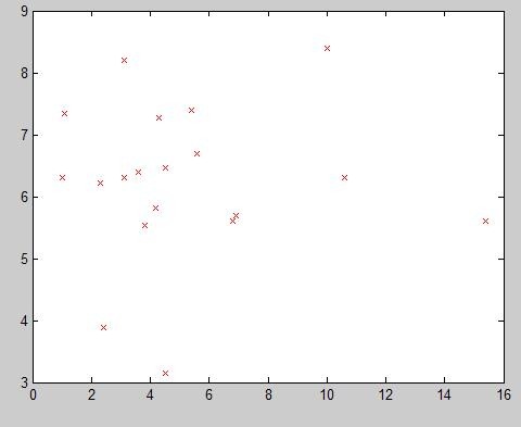
Results: Of the 19 patients, there were 5 men and 14 women. The median age at diagnosis of RF was 50 years (range 42-64 years). The most frequent presenting symptoms were back or abdominal pain, weakness, weight loss, oligoanuria, arterial hypertension and mild fever. The duration of symptoms before diagnosis ranged from 6 to 18 months. At presentation all patients had active disease, presenting renal dysfunction with a median serum creatinine of 5.18 mg/dl (range 1-15.4 mg/dl). Most of the patients had moderate bilateral hydronephrosis (2nd degree hydronephrosis). In our study, all patients had excellent prognosis, with full recovery of renal function in 78% of cases (15 patients). The fractal dimension of the fibrosis mass contour correlates with level of renal function impairment. Even more, the fractal dimension seems to slightly variate between CT evaluations (1.30 +/- 0.1), suggesting a non aggressive pattern of extension of the fibrotic mass characteristic for benign lesions.
Conclusions: The imaging parameters did not predict the disease severity, except the increase in fractal dimension of fibrosis surface area. Efficacy of bilateral ureteric stenting in improving renal function is limited in most of the cases. Dispite the level of renal function impairment at admission, full recovery can be achieved after bilateral ureteric stenting/nephrostomy and ureterolisis.
Figures


References
-
- Resnick MI, Kursh ED. Campbell's Urology . Philadelphia: WB Saunders; 1998. Extrinsic obstruction of the ureter; pp. 387–419.
-
- Ormond JK. Bilateral ureteral obstruction due to envelopment and compression by an inflammatory retroperitoneal process. J Urol. 1948;59:1072–1079. - PubMed
-
- Katz R, Golijanin D, Pode D, Shapiro A. Primary and postoperative retroperitoneal fibrosis – experience with 18 cases. Urology. 2002;60:780–783. - PubMed
-
- Khan AN, Chandramohan M. Retroperitoneal fibrosis. eMedicine .
-
- Mitchinson MJ, Withycombe JF. The response of idiopathic retroperitoneal fibrosis to corticosteroids. Br J Urol. 1971;43:44–49. - PubMed
Publication types
MeSH terms
LinkOut - more resources
Full Text Sources
Research Materials