. 2010 Mar 19:5:8.
doi: 10.1186/1749-8104-5-8.

Cortico-cerebral histogenesis in the opossum Monodelphis domestica: generation of a hexalaminar neocortex in the absence of a basal proliferative compartment

Affiliations

Cortico-cerebral histogenesis in the opossum Monodelphis domestica: generation of a hexalaminar neocortex in the absence of a basal proliferative compartment

Elisa Puzzolo et al. Neural Dev. .

Abstract

Background: The metatherian Monodelphis domestica, commonly known as the South-American short-tailed opossum, is an appealing animal model for developmental studies on cortico-cerebral development. Given its phylogenetic position, it can help in tracing evolutionary origins of key traits peculiar to the eutherian central nervous system. The capability of its pup to regenerate damaged cortico-spinal connections makes it an ideal substrate for regenerative studies. Recent sequencing of its genome and the ex utero accessibility of its developing cerebral cortex further enhance its experimental interest. However, at the moment, a comprehensive cellular and molecular characterization of its cortical development is missing.

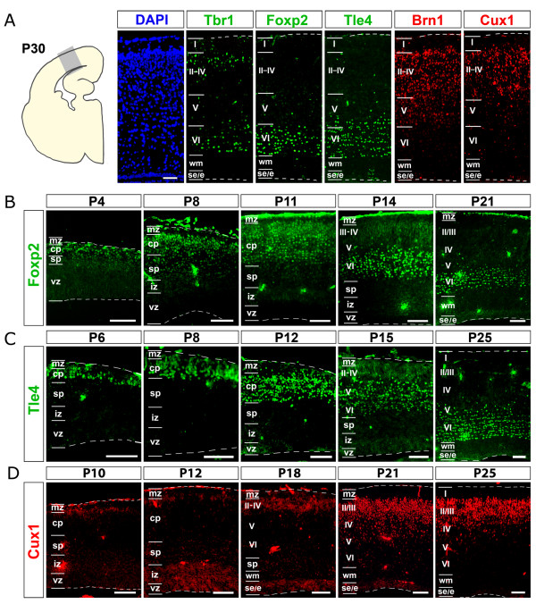
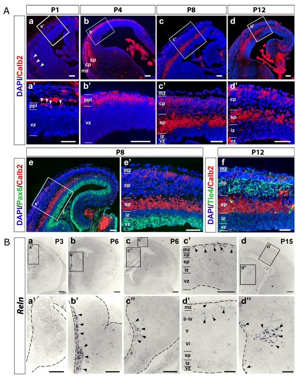
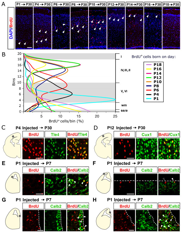
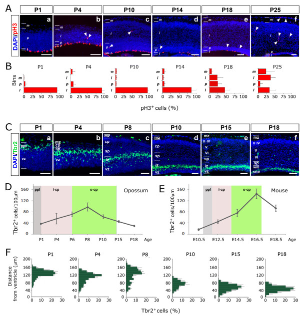
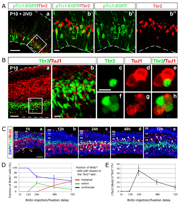
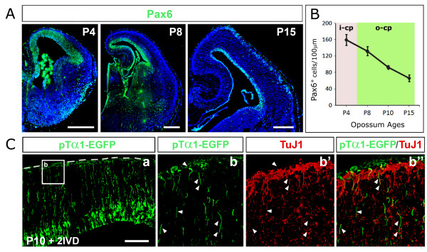
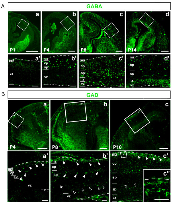
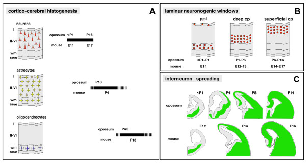
Results: A systematic analysis of opossum cortico-cerebral development was performed, including: origin of cortical neurons; migration of these neurons from their birthplaces to their final layer destinations; and molecular differentiation of distinct neocortical laminae. We observed that opossum projection neurons and interneurons are generated by pallial and subpallial precursors, respectively, similar to rodents. A six-layered cortex with a eutherian-like molecular profile is laid down, according to the inside-out rule. However, neocortical projection neurons are generated by apical neural precursors and almost no basal progenitors may be found in the neuronogenic neopallial primordium. In the opossum neocortex, Tbr2, the hallmark of eutherian basal progenitors, is transiently expressed by postmitotic progenies of apical precursors prior to the activation of more mature neuronal markers.

Conclusions: The neocortical developmental program predates Eutheria-Methatheria branching. However, in metatherians, unlike eutherians, a basal proliferative compartment is not needed for the formation of a six-layered neuronal blueprint.


Figures

Figure 1
Figure 1
Expression profiles of layer-specific markers in the opossum neocortex. (A) DAPI staining and Tbr1, Foxp2, Tle4, Brn1 and Cux1 immunofluorescence, on adjacent P30 mid-frontal neocortical sections. (B-D) Time course immunoprofiling of Foxp2, Tle4 and Cux1 on mid-frontal neocortical sections, from P4 to P25. Abbreviations: I, II, III, IV, V, VI refer to cortical layers; cp, cortical plate; e, ependyma; iz, intermediate zone; mz, marginal zone; ppl, preplate; se, subependymal zone; sp, subplate; vz, ventricular zone; wm, white matter. Scale bars: 100 μm.
Figure 2
Figure 2
Dynamics of Calretinin and Reelin expression in the developing opossum telencephalon. (Aa-d) Time course immunoprofiling of Calretinin (Calb2) from P1 to P12. (Aa'-d') Magnifications of boxed areas in (Aa-d). (Ae-f) Comparisons among distributions of Calretinin, Pax6 (Ae,e') and Tle4 (Af). (B) In situ hybridization of Reelin (Reln) mRNA on coronal sections of P3, P6 and P15 opossum telencephalons. (Ba'-d") are magnifications of boxed areas in (Ba-d). Abbreviations: II-IV, V, VI refer to cortical layers; cp, cortical plate; iz, intermediate zone; mz, marginal zone; ppl, preplate; sp, subplate; vz, ventricular zone. Scale bars: 100 μm. Arrowheads in A point to Calb2+ cells within the ppl (a,a'); arrowheads in B point to Reln+ neurons at around the septo-pallial border (b,b'), in the neocortical marginal zone (c,c'), the cortical hem (c,c",d,d"), the neocortical outer cortical plate (d,d').
Figure 3
Figure 3
Bromodeoxyuridine birthdating of opossum neocortical neurons. (A) BrdU immunoprofiling of mid-frontal neocortical sections from opossums injected with a single pulse of BrdU at the ages of P1, P4, P6, P8, P10, P12, P14, P16 and P18, and fixed at P30. Arrowheads point to grey matter BrdU+ cells. (B) Diagramatic representation of BrdU+ cells sampled in (A): the cortical wall is divided into 20 equally spaced bins, numbered from ventricular to marginal; radial extension of cortical laminae is indicated by white/grey shading; plots representing percentages of BrdU+ cells falling into each bin, for each injection time. (C-H) Colocalization of layer-specific markers, Tle4 (C), Cux1 (D), Calb2 (E-H), with BrdU injected at P4 (C), P12 (D) and P7 (E-H), respectively. Solid arrowheads in (E-H) point to Calb2+/BrdU+ cells; empty arrowheads in (F, H) point to Calb2-/BrdU+ cells. Abbreviations: I, II, III, IV, V, VI refer to cortical layers; e, ependyma; se, subependymal zone; wm, white matter. Scale bars: 100 μm in (A); 40 μm in (C-H).
Figure 4
Figure 4
Dynamics of phospho-histone3 and Tbr2 expression in the developing opossum cortex. (A) Phospho-histone3 (pH3) immunoprofiling of mid-frontal neocortical sections of P1 to P25 opossums. Immunopositive cells are prevalently aligned near the ventricle; solid arrowheads point to rare abventricular pH3+ mitotic cells. (B) Diagrammatic representation of pH3+ cells sampled in (A). (C) Tbr2 immunoprofiling of mid-frontal neocortical sections of P1 to P18 opossums. (D-F). Linear densities (D) and radial distributions (F) of Tbr2+ cells sampled in (C). (E) Linear densities of Tbr2+ cells in the mouse. Grey, pink and green shading in (D, E) demarcate peak neuronogenesis windows for primordial plexiform layer (ppl), inner cortical plate (i-cp) and outer cortical plate (o-cp), respectively. Abbreviations: II-IV, V, VI refer to cortical layers; cp, cortical plate; e, ependyma; iz, intermediate zone; mz, marginal zone; ppl, preplate; se, subependymal zone; sp, subplate; vz, ventricular zone; wm, white matter. Scale bars: 100 μm in (A, C).
Figure 5
Figure 5
Origin and fate of Tbr2+ cells. (A, B) Confocal Tbr2/enhanced green fluorescent protein (EGFP) (Aa-b") and Tbr2/β-tubulin (Ba, b) immunoprofiling of opossum cerebral cortex, dissected out at P10, acutely electroporated with a pTα1-EGFP plasmid and kept in in vitro culture for 48 h. Magnifications of boxed areas in (Aa) and (Ba) are shown in (Ab-b") and (Bb), respectively. (Bc-h) Colocalization of Tbr2 and β-tubulin on acutely dissociated cells from P10 opossum cortex. (C) Confocal Tbr2/BrdU immunoprofiling of neocortical coronal sections from opossum pups, pulsed with BrdU at P6 and sacrificed after different times: 1 h, 12 h, 24 h, 48 h and 72 h. Arrowheads in (Cc-e) point to cells immunoreactive for both BrdU and Tbr2. (D) Relative radial distribution of BrdU+ cells sampled in (C) compared to the Tbr2+ belt. (E) Time course of percentages of Tbr2+cells sampled in (C) also immunoreactive for BrdU. Abbreviations: cp, cortical plate; iz, intermediate zone; mz, marginal zone; sp, subplate; vz, ventricular zone. Scale bars: 100 μm in (A-C); 20 μm in (Ab, Bb, c).
Figure 6
Figure 6
Analysis of the apical progenitor compartment. (A) Pax6 immunoprofiling of a selection of coronal sections from P4 to P15 opossum telencephalons. (B) Linear densities of Pax6+ cells sampled in (A). (C) EGFP/β-tubulin immunoprofiling of opossum cerebral cortex, dissected out at P10, acutely electroporated with a pTα1-EGFP plasmid and kept in in vitro culture for 48 h. Magnifications of the boxed area in (Ca) are shown in (Cb-b"). Arrowheads in (Cb-b") point to pial processes of electroporated cells, immunoreactive for EGFP, but not for β-tubulin. Abbreviations: i-cp, inner cortical plate; o-cp, outer cortical plate. Scale bars: 400 μm in (A); 100 μm in (C).
Figure 7
Figure 7
Distribution of GABA and glutamate-decarboxylase immunoreactivity in developing opossum cortices. (Aa-d) Distribution of GABA+ cells on frontal sections of P1 to P14 cortices. (Aa'-d') Magnifications of boxed regions in (Aa-d). (Ba-c) Distribution of glutamate-decarboxylase (GAD)+ cells on frontal sections of P4, P8 and P10 cortices. (Ba'-c') Magnifications of boxed regions in (Ba-c). GAD+ cells are evident in the marginal zone and the subplate since P4 (Ba',b', solid arrowheads). At P8, an additional reactivity is detectable in proliferative layers (Bb', empty arrowheads). Finally, at P10, both marginal zone and periventricular GAD signals are strengthened (Bc',c") and additional GAD+ cells are present throughout the cortical plate (Bc'). Abbreviations: cp, cortical plate; iz, intermediate zone; mz, marginal zone; sp, subplate; vz, ventricular zone. Scale bars: 400 μm in (Aa-d,Ba-c); 50 μm in (Aa'-d', Ba'-c").
Figure 8
Figure 8
Opossum and mouse cortico-cerebral histogenesis: a comparison. (A) Radial distribution and approximate temporal generation windows of neurons, astrocytes and oligodendrocytes in the opossum and mouse cerebral cortex. (B) Peak generation times for primordial plexiform layer, deep cortical plate and superficial cortical plate in opossum and mouse. (C) Temporal profile of interneurons spreading in the developing cerebral cortex of opossum and mouse. Abbreviations: I, II-VI refer to cortical layers; cp, cortical plate; e, ependyma; ppl, primordial plexiform layer; se, subependymal zone; wm, white matter.

Similar articles

Cited by

References

    1. Ji Q, Luo ZX, Yuan CX, Wible JR, Zhang JP, Georgi JA. The earliest known eutherian mammal. Nature. 2002;416:816–822. doi: 10.1038/416816a. - DOI - PubMed
    1. Kumar S, Hedges SB. A molecular timescale for vertebrate evolution. Nature. 1998;392:917–920. doi: 10.1038/31927. - DOI - PubMed
    1. Woodburne M, Rich T, Springer MS. The evolution of tribospheny and the antiquity of mammalian clades. Mol Phylogenet Evol. 2003;28:360–385. doi: 10.1016/S1055-7903(03)00113-1. - DOI - PubMed
    1. Nicholls JG, Saunders N. Regeneration of immature mammalian spinal cord after injury. Trends Neurosci. 1996;19:229–234. doi: 10.1016/0166-2236(96)10021-7. - DOI - PubMed
    1. Saunders NR, Adams E, Reader M, Møllgard K. Monodelphis domestica (gray short-tailed opossum): an accessible model for studies of early neocortical development. Anat Embryol. 1989;180:227–236. doi: 10.1007/BF00315881. - DOI - PubMed

Publication types

MeSH terms