. 2010 Mar 19:11:187.
doi: 10.1186/1471-2164-11-187.

Antimicrobial peptide-like genes in Nasonia vitripennis: a genomic perspective

Affiliations

Antimicrobial peptide-like genes in Nasonia vitripennis: a genomic perspective

Caihuan Tian et al. BMC Genomics. .

Abstract

Background: Antimicrobial peptides (AMPs) are an essential component of innate immunity which can rapidly respond to diverse microbial pathogens. Insects, as a rich source of AMPs, attract great attention of scientists in both understanding of the basic biology of the immune system and searching molecular templates for anti-infective drug design. Despite a large number of AMPs have been identified from different insect species, little information in terms of these peptides is available from parasitic insects.

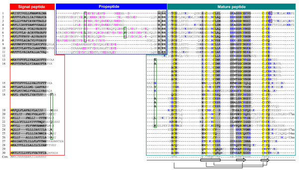
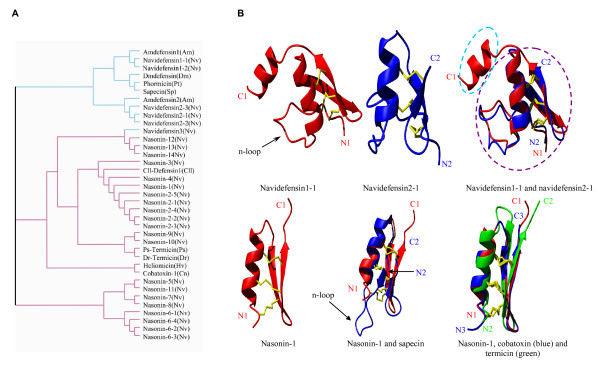
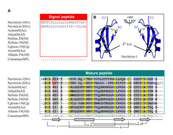
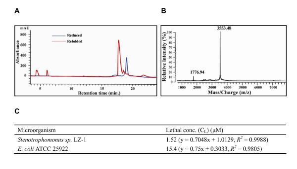
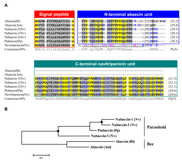
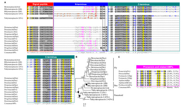
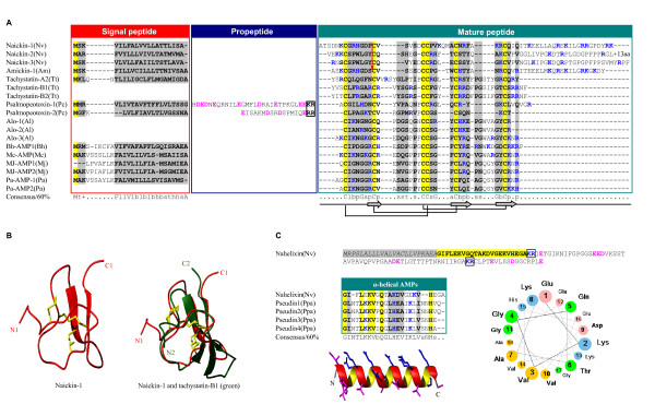
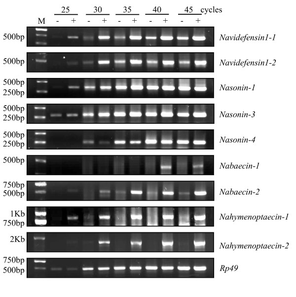
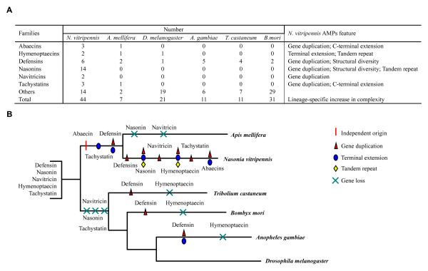
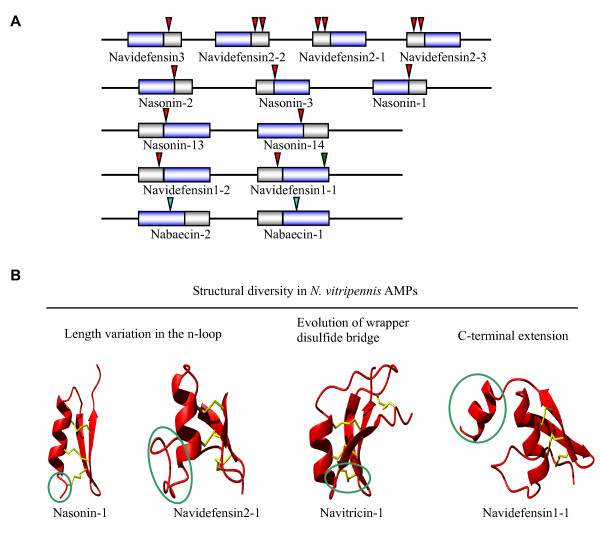
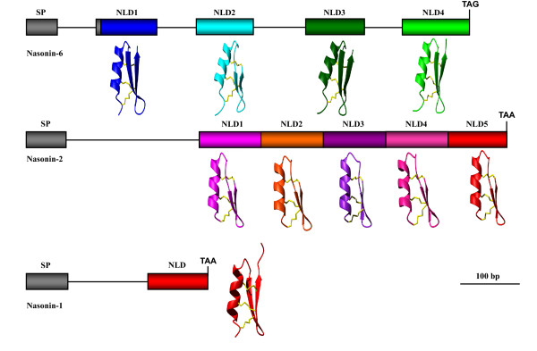
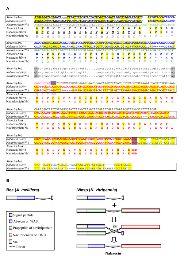
Results: By using integrated computational approaches to systemically mining the Hymenopteran parasitic wasp Nasonia vitripennis genome, we establish the first AMP repertoire whose members exhibit extensive sequence and structural diversity and can be distinguished into multiple molecular types, including insect and fungal defensin-like peptides (DLPs) with the cysteine-stabilized alpha-helical and beta-sheet (CSalphabeta) fold; Pro- or Gly-rich abaecins and hymenoptaecins; horseshoe crab tachystatin-type AMPs with the inhibitor cystine knot (ICK) fold; and a linear alpha-helical peptide. Inducible expression pattern of seven N. vitripennis AMP genes were verified, and two representative peptides were synthesized and functionally identified to be antibacterial. In comparison with Apis mellifera (Hymenoptera) and several non-Hymenopteran model insects, N. vitripennis has evolved a complex antimicrobial immune system with more genes and larger protein precursors. Three classical strategies that are likely responsible for the complexity increase have been recognized: 1) Gene duplication; 2) Exon duplication; and 3) Exon-shuffling.

Conclusion: The present study established the N. vitripennis peptidome associated with antimicrobial immunity by using a combined computational and experimental strategy. As the first AMP repertoire of a parasitic wasp, our results offer a basic platform for further studying the immunological and evolutionary significances of these newly discovered AMP-like genes in this class of insects.


Figures

Figure 1
Figure 1
Multiple sequence alignment of navidefensins, nasonins and related peptides. 1: Amdefensin1(Am); 2: Amdefensin2(Am); 3: Navidefensin1-1(Nv); 4: Navidefensin1-2(Nv); 5: Navidefensin2-1(Nv); 6: Navidefensin2-2(Nv); 7: Navidefensin2-3(Nv); 8: Navidefensin3(Nv); 9: Phormicin(Pt); 10: Sapecin(Sp); 11: Dmdefensin(Dm); 12: Heliomicin(Hv); 13: Nasonin-1(Nv); 14: Nasonin-2(Nv); 15: Nasonin-3(Nv); 16: Nasonin-4(Nv); 17: Nasonin-5(Nv); 18: Nasonin-6(Nv); 19: Nasonin-7(Nv); 20: Nasonin-8(Nv); 21: Nasonin-9(Nv); 22: Nasonin-10(Nv); 23: Nasonin-11(Nv); 24: Nasonin-12(Nv); 25: Nasonin-13(Nv); 26: Nasonin-14(Nv); 27: Cobatoxin-1(Cn); 28: Cll-Defensin1(Cll); 29: Ps-Termicin(Ps); 30: Dr-Termicin(Dr). Am: A. mellifera, Nv: N. vitripennis, Pt: Protophormia terraenovae, Sp: Sarcophaga peregrine, Dm: D. melonagaster, Hv: Heliothis virescens, Cn: Centruroides noxius, Cll: C. limpidus limpidus, Ps: Pseudacanthotermes spiniger, Dr: Drepanotermes rubriceps. Secondary structure elements (α-helix: cylinder; β-strand: arrow) and disulfide bridge connectivity are shown on the bottom of the alignment. Acidic residues in the propeptide are shown in pink and cleavage sites are boxed. Identical amino acids or constitutive replacements are shadowed in yellow and grey, respectively. Consensu/60% (Con.): - (negative), * (Ser/Thr), l (aliphatic), + (positive), t (tiny), a (aromatic), c (charged), s (small), p (polar), b (big), h (hydrophobic). Basic residues (K, R, H) are shown in blue. Residues split by phase 1 or 2 introns are boxed in green or purple. Omitted residues in carboxyl-termini are indicated by +aa.
Figure 2
Figure 2
Clustering and structure of DLPs. A. Clustering analysis of the sequences in Figure 1 by the CLUSTAL program. Cyan and purple branches respectively represent CITDs, and nasonins and related peptides. Nasonin-2 and nasonin-6 are aligned as repeated domains; B. Structural models of DLPs from N.vitripennis. Navidefensin1-1 and navidefensin2-1 modeled ab initio by I-TASSER are shown in ribbon displayed by MolMol. Superimposition of these two structures shows an extra carboxyl-terminal domain in navidefensin1-1 structure. The model structure of nasonin-1 is compared with those of sapecin, termicin and cobatoxin.
Figure 3
Figure 3
Navitricins. A. Sequence alignment of navitricins with fungal DLPs. The position of a phase 1 intron is boxed in red; B. The model structure of navitricin-1. Its fourth bridge, which links the n-loop to c-loop, is indicated by arrows. Ac: Aspergillus clavatus, Af: A. fumigatus, Nf: Neosartorya fischeri, Cg: Chaetomium globosum, Ao: A. oryzae, Afl: A. flavus.
Figure 4
Figure 4
Purification and characterization of nasonin-1. A. RP-HPLC showing retention time difference between the reduced and refolded products; B. MALDI-TOF MS of the refolded nasonin-1; C. Antibacterial activities of nasonin-1.
Figure 5
Figure 5
Abaecin-like peptides. A. Multiple sequence alignment. The propeptide sequence of navitripenicin is italized and underlined once. [0] indicates a phase 0 intron between the two residues and [?] represents unknown intron information due to the lack of genomic sequence. B. Phylogenetic tree constructed with the NJ method of MEGA 4.0. Solid circle represents gene duplication. Bi: Bombus ignites; Pp: Pteromalus puparum.
Figure 6
Figure 6
Hymenoptaecin-like peptides. A. Multiple sequence alignment. Red arrows label the position of phase 0 introns. A red "^" indicates a site in droyataecin that is corrected from TAG to GAG codon. Proline residues in amino-termini of nahymenoptaecins are bolded and shown in brown. Putative proprotein cleavage sites are boxed in black; B. Phylogenetic tree constructed with the NJ method of MEGA 4.0. Solid circle represents gene duplication; C. Comparison of pronavicin with known Pro-rich AMPs. Sequence identity with pronavicin and proline contents are shown here. Dse: D. sechellia, Dsi: D. simulans, Dpe: D. persimilis, Dps: D. pseudoobscura, Dvi: D. virilis, Dya: D. yakuba, Der: D. erecta, Dan: D. ananassae, Dgr: D. grimshawi, Dwi: D. willistoni, Mg: Myrmecia gulosa, Pa: Pyrrhocoris apterus, Bp: Bombus pascuorum.
Figure 7
Figure 7
Tachystatin-type and linear AMPs. A. Multiple sequence alignment of tachystatin-type AMPs. A red arrow represents a phase 0 intron; B. Model structure of naickin-1 and its superimposition with tachystatin-B1; C. Nahelixin. Signal peptide and mature peptides are shaded in gray and yellow, respectively. In the alignment of nahelixin with known AMPs from frog, identical residues are bolded and shadowed in yellow while conservative substitutions are in gray. Structure modeled ab initio is displayed in ribbon with hydrophobic residues in pink and hydrophilic residues in blue. The helical wheel projection shows the amphiphilic characteristics of nahelixin. Putative proprotein cleavage sites are boxed in black. Tt: Tachypleus tridentatus, Pc: Psalmopoeus cambridgei, Al: Acrocinus longimanus, Bh: Bradysia hygida, Mc: Mesembryanthemum crystallinum, Mj: Mirabilis jalapa, Pa: Phytolacca americana, Ppa: Pseudis paradoxa.
Figure 8
Figure 8
Semi-quantitative RT-PCR detecting the inducible expression of AMP genes in N. vitripennis before and after bacterial infection. M: DNA marker. -: non-challenged; +: challenged.
Figure 9
Figure 9
AMPs from N. vitripennis and other insects. A. Number and feature comparison; B. Evolutionary divergence of N. vitripennis AMPs, in which gene duplication, terminal extension, tandem repeat and gene loss events are highlighted.
Figure 10
Figure 10
Gene duplication and structural diversity of AMPs in N. vitripennis. A. Gene duplication. Chromosome fragments are shown in lines and genes coding AMPs are represented by box, in which gray one stands for signal and pro-peptide and blue one indicates mature peptide. Introns are indicated in triangles with phase 1 in red, phase 2 in green, and phase 0 in cyan; B. Structural diversity in N. vitripennis AMPs. Changes in structure relative to classical insect defensins are circled.
Figure 11
Figure 11
Exon duplication of nasonins. Peptides are indicated in boxes with color in gray for signal peptide (SP) and in other colors for NLDs. Lines mean the phase 1 introns between exons. Corresponding model structures of each domain are displayed in ribbon with different colors.
Figure 12
Figure 12
A proposed exon shuffling mechanism for origin of nabaecin. A. Sequence alignment of abaecin, nabaecin-3 and navitripenicin. Identical nucleotides and amino acids are shadowed in yellow and signal peptides are italized and underlined once. Alignable sequences are bolded and the corresponding nucleotides are boxed. Intron splice sites are showed in gray and omitted sequences are displayed as dots. Stop codons are shadowed in dark gray; B. The exon-shuffing model (for detailed description, see the text). Color codes are the same with those in Figure 12A.

Similar articles

Cited by

References

    1. Ezekowitz RA, Hoffmann JA. Innate immunity. Totowa, New Jersey: Human press; 2003.
    1. Hale JD, Hancock RE. Alternative mechanisms of action of cationic antimicrobial peptides on bacteria. Expert Rev Anti Infect Ther. 2007;5:951–959. doi: 10.1586/14787210.5.6.951. - DOI - PubMed
    1. Brogden KA. Antimicrobial peptides: pore formers or metabolic inhibitors in bacteria? Nat Rev Microbiol. 2005;3:238–250. doi: 10.1038/nrmicro1098. - DOI - PubMed
    1. Jenssen H, Hamill P, Hancock RE. Peptide antimicrobial agents. Clin Microbiol Rev. 2006;19:491–511. doi: 10.1128/CMR.00056-05. - DOI - PMC - PubMed
    1. Brown KL, Hancock RE. Cationic host defense (antimicrobial) peptides. Curr Opin Immunol. 2006;18:24–30. doi: 10.1016/j.coi.2005.11.004. - DOI - PubMed

Publication types

Substances