. 2010 Jun 1;342(1):1-10.
doi: 10.1016/j.ydbio.2010.03.005. Epub 2010 Mar 17.

Regulation of EGFR and Notch signaling by distinct isoforms of D-cbl during Drosophila development

Affiliations

Regulation of EGFR and Notch signaling by distinct isoforms of D-cbl during Drosophila development

Yuan Wang et al. Dev Biol. .

Abstract

Cells receive and interpret extracellular signals to regulate cellular responses such as proliferation, cell survival and differentiation. However, proper inactivation of these signals is critical for appropriate homeostasis. Cbl proteins are E3-ubiquitin ligases that restrict receptor tyrosine kinase (RTK) signaling, most notably EGFR (Epidermal Growth Factor Receptor), via the endocytic pathway. Consistently, many mutant phenotypes of Drosophila cbl (D-cbl) are due to inappropriate activation of EGFR signaling. However, not all D-cbl phenotypes can be explained by increased EGFR activity. Here, we report that D-Cbl also negatively regulates Notch activity during eye and wing development. D-cbl produces two isoforms by alternative splicing. The long isoform, D-CblL, regulates the EGFR. We found that the short isoform, D-CblS, preferentially restricts Notch signaling. Specifically, our data imply that D-CblS controls the activity of the Notch ligand Delta. Taken together, these data suggest that D-Cbl controls the EGFR and Notch/Delta signaling pathways through production of two alternatively spliced isoforms during development in Drosophila.


Figures

Figure 1
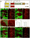
Figure 1. Increased R8 spacing in D-cbl clones is Notch-dependent
(A) Domain structure of the long and short isoforms of D-Cbl. The relative locations of the non-sense mutation in D-cblK26 and the missense mutations in D-cbl7 are indicated. TKB - tyrosine kinase binding domain; L – Linker; RING - RING E3 ubiquitin ligase; UBA - ubiquitin-associated domain. (B,B′) Increased spacing between R8 photoreceptor cells in D-cblK26 mutant clones. Anti-Senseless (Sens) labeling is shown in red to mark R8 cells. Anterior is to the left. Absence of GFP marks D-cbl mutant clones outlined by white lines in (B′). White triangles mark the position of the morphogenetic furrow. Genotype: ey-Flp; D-cblK26 FRT80/P[ubi-GFP] FRT80. (C,C′) Heterozygosity of Notch (N) normalizes R8 spacing in D-cblK26 mutant clones. Mutant clones are outlined by white lines in (C′). White triangles mark the position of the morphogenetic furrow. Genotype: ey-Flp/Df(1)N8; D-cblK26 FRT80/P[ubi-GFP] FRT80. A similar result was obtained with N264-39 (data not shown). (D,D′) Increased ommatidial spacing in D-cblK26 clones in 42 hours APF pupal eye discs labeled with Elav (marks photoreceptor neurons or R cells) in red. The D-cbl clone is marked by absence of GFP and outlined by a white line in (D′). Genotype as in (B). (E) Summary of the analysis of R8 spacing. The distances between R8 cells in the MF and posterior to the MF in eye imaginal discs of the indicated genotypes are measured by Axiovision 6 program with the average length per R8 pairs in μM. A total of 100 R8 pairs from 10 different discs were measured for each data point. (F,F′) argosΔ7 and (G,G′) gap123-9s mutant clones do not affect R8 spacing as determined by Sens labeling in red. Mutant clones are outlined by white lines in (F′,G′). White triangles mark the position of the morphogenetic furrow. Genotypes: ey-Flp; argosΔ7 or gap123-9s FRT80/P[ubi-GFP] FRT80.
Figure 2
Figure 2. Partial rescue of R7 and cone cell defects in egfr+/− and N+/− background
Analysis of R7, photoreceptors (R cells) (A-A‴) and cone cells (B,B′) in D-cblK26 clones in heterozygous Notch (N) background by Pros/Elav (red/blue) double labelings (A-A‴) and Cut (red) labelings (B,B′) in 42 hrs APF pupal eye imaginal discs. Mutant clones are outlined by white lines in A′-A‴ and B′. Compared to D-cblK26 clones alone (Fig. 7A,B), R7 and cone cells are significantly reduced in N/+;D-cbl mutant ommatidia (see quantification in D). Genotype: ey-Flp/Df(1)N8; D-cblK26 FRT80/P[ubi-GFP] FRT80. (C,C′) Anti-Pros (red) and anti-Elav (blue) double labeling of D-cblK26 mosaics heterozygous for egfr in 42 hours APF eye disc. Mutant clones are outlined by white lines in C′. Genotype: ey-Flp; egfrf2/+; D-cblK26 FRT80/P[ubi-GFP] FRT80. (D) Summary of the total number of R7 and cone cells in 42 hrs APF pupal eye imaginal discs of the indicated genotypes as determined by Anti-Pros and Elav staining. A total of 35 ommatidia were counted for each genotype. (E) List of cell type-specific markers and the signaling pathways involved in specification of the cell types listed.
Figure 3
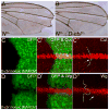
Figure 3. D-Cbl antagonizes Notch signaling
(A,B) Adult wing of a Notch heterozygote (A) and a trans-heterozygote of Notch and D-cblK26 (B). The Notch allele used is Df(1)N8. (C-C″) Anti-Cut (red) labeling of D-cblK26 mosaics in wing imaginal discs induced by the MARCM system. Homozygous mutant clones are marked by GFP (C,C′). The white line in (C″) marks the clonal boundary. The arrow points to non-autonomous increase of Cut labeling. Genotype: hs-Flp actin-Gal4 UAS-GFP; tub-Gal80 FRT80/D-cblK26 FRT80. (D-D″) Anti-Wingless (Wg) (red) labeling of D-cblK26 mosaics in wing imaginal discs induced by the MARCM system. Homozygous mutant clones are marked by GFP (D,D′). The white line in (D″) marks the clonal boundary. Note the increased Wg immunolabeling in D-cbl mutant tissue (arrow in D″). Genotype in (C) and (D): hs-Flp actin-Gal4 UAS-GFP; tub-Gal80 FRT80/D-cblK26 FRT80.
Figure 4
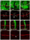
Figure 4. D-CblS suppresses the Notch target gene Cut
Wing imaginal discs were labeled for Cut as Notch activity marker in red. Clones over-expressing D-cblL (A,A′), D-cblS (B,B′) and EGFRDN (C,C′) were obtained by crossing the corresponding UAS lines to hs-Flp; tub<GFP<Gal4 flies, and are negatively marked by the absence of GFP. The top panels of each experiment are the combined GFP and Cut channels, the (′) panels are the Cut labelings only. Expression of D-cblS reduced expression of Cut (B,B′, arrow). Induction of D-cblL (A,A′) and EGFRDN (C,C′) did not affect Cut expression. White lines mark the clone boundaries. Expression of D-CblS (E,E′) specifically along the AP boundary of wing discs by dpp-Gal4, positively marked by GFP, strongly suppresses Cut expression (arrow in E′). Expression of D-cblL (D,D′) and EGFRDN (F,F′) have no effect in this assay. The UAS-D-cblL (line A18) and UAS-D-cblS (line A1) transgenes were used.
Figure 5
Figure 5. D-CblL negatively regulates EGFR activity
Heat shock-induced expression of D-cblL (marked by absence of GFP) reduces pTyr labeling (in red) (A,A′) and dpERK protein (red) levels (C,C′) in 3rd instar eye discs. The top panels of each experiment are the combined GFP and pTyr/dpERK channels, the (′) panels are the pTyr/dpERK labelings only. Heat shock-induced clones expressing D-cblS do not affect pTyr (B,B′) and dpERK levels (D,D′). The white triangle marks the position of the MF. White lines in (A′,B′,C′,D′) mark the clone boundaries. The arrows in (D′) point to dpERK positive cells in D-cblS expressing clones. The UAS-D-cblL (line A18) and UAS-D-cblS (line A1) transgenes were used.
Figure 6
Figure 6. D-CblS negatively regulates Delta
(A,B) Overexpression of Delta (Dl) (A,A′) and Serrate (Ser) (B,B′) along the AP boundary using dpp-Gal4 induces ectopic Cut expression in the dorsal or ventral part of the dpp-expression domain. GFP marks the dpp expression domain. (C,D) Co-expression with D-cblS suppresses Delta-induced Cut expression (C,C′), but has little effect on Ser-induced Cut expression (D,D′). GFP marks the dpp expression domain. (E,F) Accumulation of Delta protein in D-cbl7 mutant clones posterior to the MF (E,E′) and in the wing disc (E,F). D-cbl7 affects the RING E3-ubiquitin ligase domain of D-Cbl (Fig. 1A). Delta protein does not accumulate in D-cbl clones in regions where Delta is normally not present. Clones are marked by the absence of GFP and outlined by white lines in E′ and F′ (see arrows). Genotypes: (E,E′) ey-Flp; D-cbl7 FRT80/P[ubi-GFP] FRT80. (F,F′) hs-Flp; D-cbl7 FRT80/P[ubi-GFP] FRT80.
Figure 7
Figure 7. Distinct rescue of the D-cbl over-recruitment phenotype by expression of the D-cbl isoforms
Shown are pupal (42h APF) eye discs labeled with anti-Pros/Elav antibodies (red/blue) for R7/photoreceptor (R) cells and anti-Cut antibody (red) for cone cells. D-cbl mutant clones are positively labeled by GFP due to the MARCM technique (outlined by white lines) and express either no transgene (A,A,B,B′), D-cblL (C,C,D,D′) or D-cblS (E,EF,F′). GFP-negative cells are non-mutant (wild-type or heterozygous) and do not express any transgene. Arrows point to representative examples of the phenotypes in the mutant clones as explained in the text. The null allele D-cblK26, and the transgenes UAS-D-cblL (line A18) and UAS-D-cblS (line A1) transgenes were used. Genotypes: (A,B) hs-Flp actin-Gal4 UAS-GFP; D-cblK26 FRT80/tub-Gal80 FRT80; (C,D) hs-Flp actin-Gal4 UAS-GFP; UAS-D-cblL/+; D-cblK26 FRT80/tub-Gal80 FRT80; (E,F) hs-Flp actin-Gal4 UAS-GFP; UAS-D-cblS/+; D-cblK26 FRT80/tub-Gal80 FRT80.
Figure 8
Figure 8. Summary of D-Cbl activities
Shown are the characterized activities of D-CblL and D-CblS in this report and by (Wang et al., 2008b) during eye and wing development. The dotted line indicates a potential minor activity of D-CblS towards EGFR.

Similar articles

Cited by

References

    1. Baker NE, Zitron AE. Drosophila eye development: Notch and Delta amplify a neurogenic pattern conferred on the morphogenetic furrow by scabrous. Mech Dev. 1995;49:173–189. - PubMed
    1. Baonza A, Casci T, Freeman M. A primary role for the epidermal growth factor receptor in ommatidial spacing in the Drosophila eye. Curr Biol. 2001;11:396–404. - PubMed
    1. Bergmann A, Agapite J, McCall K, Steller H. The Drosophila gene hid is a direct molecular target of Ras-dependent survival signaling. Cell. 1998;95:331–341. - PubMed
    1. Cagan RL, Ready DF. The emergence of order in the Drosophila pupal retina. Dev Biol. 1989;136:346–362. - PubMed
    1. Casci T, Vinos J, Freeman M. Sprouty, an intracellular inhibitor of Ras signaling. Cell. 1999;96:655–665. - PubMed

Publication types

MeSH terms