Orosensory deprivation alters taste-elicited c-Fos expression in the parabrachial nucleus of neonatal rats
- PMID: 20302893
- PMCID: PMC2884291
- DOI: 10.1016/j.neures.2010.03.007
Orosensory deprivation alters taste-elicited c-Fos expression in the parabrachial nucleus of neonatal rats
Abstract
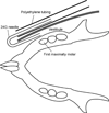
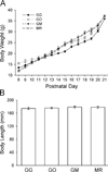
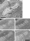
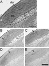
In the present study we examined the effects of neonatal orosensory deprivation on taste-elicited gustatory activity in the rat parabrachial nucleus (PBN) using the functional anatomical marker c-Fos. Animals in three groups (GG, GO and GM) received gastric cannula implantation surgery on postnatal day 9 (P9). Animals in the fourth group (MR) did not receive any surgery. GG rats were fed by infusion of artificial milk directly into the stomach. GO rats were fed by intraoral infusion of artificial milk. GM and MR rats were reared by their mother with free access to mother's milk, water and rat chow. Rats from all groups were similar in body weight and length by P21. On P21 rats in all groups were intraorally presented with 0.5M sucrose solution and the brains were extracted and processed for c-Fos immunohistochemistry. Taste-elicited c-Fos expression in both the gustatory waist area, and the external lateral subnucleus of the PBN in rats in the GG group was significantly more robust than in the other three groups. These findings suggest a substantial alteration in orosensory-evoked neuronal response in this nucleus, due to sensory or motor deprivation during a critical developmental stage.
Copyright 2010 Elsevier Ireland Ltd and the Japan Neuroscience Society. All rights reserved.
Figures


Similar articles
-
Expression of Fos during sham sucrose intake in rats with central gustatory lesions.Am J Physiol Regul Integr Comp Physiol. 2008 Sep;295(3):R751-63. doi: 10.1152/ajpregu.90344.2008. Epub 2008 Jul 16. Am J Physiol Regul Integr Comp Physiol. 2008. PMID: 18635449 Free PMC article.
-
c-Fos expression in the parabrachial nucleus after ingestion of sodium chloride in the rat.Neuroreport. 1993 Sep 10;4(11):1223-6. doi: 10.1097/00001756-199309000-00003. Neuroreport. 1993. PMID: 8219018
-
Effects of gustatory nerve transection and regeneration on quinine-stimulated Fos-like immunoreactivity in the parabrachial nucleus of the rat.J Comp Neurol. 2003 Oct 13;465(2):296-308. doi: 10.1002/cne.10851. J Comp Neurol. 2003. PMID: 12949788
-
C-fos expression in the brainstem after voluntary ingestion of sucrose in the rat.Neurobiology (Bp). 1996;4(1-2):85-102. Neurobiology (Bp). 1996. PMID: 9116697
-
The parabrachial nucleus and conditioned taste aversion.Brain Res Bull. 1999 Feb;48(3):239-54. doi: 10.1016/s0361-9230(98)00173-7. Brain Res Bull. 1999. PMID: 10229331 Review.
Cited by
-
Subnuclear organization of parabrachial efferents to the thalamus, amygdala and lateral hypothalamus in C57BL/6J mice: a quantitative retrograde double labeling study.Neuroscience. 2010 Nov 24;171(1):351-65. doi: 10.1016/j.neuroscience.2010.08.026. Epub 2010 Sep 9. Neuroscience. 2010. PMID: 20832453 Free PMC article.
-
Neural Mechanisms That Underlie Angina-Induced Referred Pain in the Trigeminal Nerve Territory: A c-Fos Study in Rats.ISRN Pain. 2013 Jul 28;2013:671503. doi: 10.1155/2013/671503. eCollection 2013. ISRN Pain. 2013. PMID: 27335881 Free PMC article.
-
MSG-Evoked c-Fos Activity in the Nucleus of the Solitary Tract Is Dependent upon Fluid Delivery and Stimulation Parameters.Chem Senses. 2016 Mar;41(3):211-20. doi: 10.1093/chemse/bjv082. Epub 2016 Jan 13. Chem Senses. 2016. PMID: 26762887 Free PMC article.
References
-
- Anseloni VC, Ren K, Dubner R, Ennis M. A brainstem substrate for analgesia elicited by intraoral sucrose. Neuroscience. 2005;133:231–243. - PubMed
-
- Ayano R, Tamura F, Ohtsuka Y, Mukai Y. The development of normal feeding and swallowing: Showa University study of the feeding function. Int. J. Orofacial Myol. 2000;26:24–32. - PubMed
-
- Baird JP, Travers SP, Travers JB. Integration of gastric distension and gustatory responses in the parabrachial nucleus. Am. J. Physiol. Regul. Integr. Comp. Physiol. 2001;281:R1581–R1593. - PubMed
-
- Barlow LA. Gustatory system development. In: Finger TE, Silver WL, Restrepo D, editors. The Neurobiology of Taste and Smell. New York: Wiley-Liss; 2000. pp. 395–422.
-
- Bernard JF, Huang GF, Besson JM. The parabrachial area: electrophysiological evidence for an involvement in visceral nociceptive processes. J. Neurophysiol. 1994;71:1646–1660. - PubMed
Publication types
MeSH terms
Substances
Grants and funding
LinkOut - more resources
Full Text Sources

