. 2010 Apr 2;141(1):154-165.
doi: 10.1016/j.cell.2010.02.037. Epub 2010 Mar 18.

Molecular and cellular approaches for diversifying and extending optogenetics

Affiliations

Molecular and cellular approaches for diversifying and extending optogenetics

Viviana Gradinaru et al. Cell. .

Abstract

Optogenetic technologies employ light to control biological processes within targeted cells in vivo with high temporal precision. Here, we show that application of molecular trafficking principles can expand the optogenetic repertoire along several long-sought dimensions. Subcellular and transcellular trafficking strategies now permit (1) optical regulation at the far-red/infrared border and extension of optogenetic control across the entire visible spectrum, (2) increased potency of optical inhibition without increased light power requirement (nanoampere-scale chloride-mediated photocurrents that maintain the light sensitivity and reversible, step-like kinetic stability of earlier tools), and (3) generalizable strategies for targeting cells based not only on genetic identity, but also on morphology and tissue topology, to allow versatile targeting when promoters are not known or in genetically intractable organisms. Together, these results illustrate use of cell-biological principles to enable expansion of the versatile fast optogenetic technologies suitable for intact-systems biology and behavior.


Figures

Figure 1
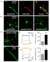
Figure 1. Multiple Trafficking Modules for Microbial Opsin Function in Mammalian Neurons
(A) Hippocampal neurons showing wild-type NpHR-EYFP aggregation in the ER. NpHR (green) and ER (red; immunostaining of the KDEL marker) are also shown in overlay (yellow) to illustrate colocalization of NpHR aggregates and ER (arrows, ER; arrowheads, ER aggregates). (B) Multistep modification of wild-type NpHR (left) rescues membrane expression. Addition of the K-channel ER export motif (eNpHR2.0, middle) and trafficking signal (eNpHR3.0, right) respectively reduced ER aggregation and improved membrane expression (particularly in neuronal processes for 3.0). (C) Membrane expression enabled in processes for eNpHR3.0 (confocal images showing membrane-localized EYFP fluorescence in the soma [top] and dendrite [inset, bottom]). (D) Representative traces (left) showing photocurrents in cells virally transduced with eNpHR3.0 (black) and eNpHR2.0 (gray). Summary plot (right) showing average photocurrent levels in cells expressing eNpHR3.0 (747.2 ± 93.9 pA) and eNpHR2.0 (214.1 ± 24.7 pA; unpaired t test p < 0.0005; n = 10). Values plotted are mean ± SEM. Membrane input resistance was similar for all neurons patched (eNpHR2.0: 193.1 ± 36.6 MΩ; eNpHR3.0: 151.6 ± 28.5 MU; unpaired t test p = 0.37). Light delivery (593 nm) is indicated by the yellow bar. (E) Representative traces (left) showing voltage traces in cells virally transduced with eNpHR3.0 (black) and eNpHR2.0 (gray). Summary plot (right) showing average hyperpolarization levels in cells expressing eNpHR3.0 (101.0 ± 24.7 mV) and eNpHR2.0 (57.2 ± 6.8 mV; unpaired t test p < 0.0005; n = 10). See also Figure S1.
Figure 2
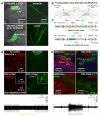
Figure 2. Trafficking-Enhanced Projection Targeting and Topological Targeting In Vivo
(A) Lentiviral delivery of eNpHR3.1 (shorter version of eNpHR3.0 with the N-terminal signal peptide removed; Experimental Procedures) driven by the CaMKIIα promoter led to expression in CA1 pyramidal neurons and dendrites (top; arrowhead indicates CA1), as well as in axon terminals (bottom; arrowhead indicates subiculum). (B) Transcellular gene activation by wheat germ agglutinin (WGA)-Cre fusion. The schematic depicts two injection sites (one with WGA-Cre fusion gene and another with Cre-dependent opsin virus) and long-range projections; Cre can be transcellularly delivered from transduced cells (red) to activate distant gene expression only in connected neurons that have received the Cre-dependent virus (green), not in others (gray). (C) Construct design for the WGA-Cre (top) and Cre-dependent (bottom) AAV vectors. WGA and Cre genes are both optimized with mammalian codons. (D) Injection of AAV carrying the EF1α::mCherry-IRES-WGA-Cre cassette in the rat primary somatosensory cortex (S1) led to mCherry fluorescence in neuronal somata at the injection site (top left), but, as expected, not in distant motor cortex (M1, bottom left). Injection of Cre-inducible AAV5-EF1α::eNpHR3.0-EYFP into the ipsilateral motor cortex (M1) led to opsin expression (EYFP fluorescence) in M1 somata (bottom right), but, as expected, in S1, eNpHR3.0-EYFP was seen only in projecting axon terminals from these M1 cells (dispersed green puncta), not in somata (top right). Optrode recordings in vivo from the M1 site (560 nm light; bottom trace). (E) The two dentate gyri were injected with AAV2-EF1a-mCherry-IRES-WGA-Cre (left) and AAV8-EF1a-DIO-ChR2-EYFP (right). Lower panels show high-magnification images of boxed regions above. Cells transduced with WGA-Cre expressed mCherry as expected (red), almost entirely within the granule cell layer (GCL) delineated by DAPI fluorescence (blue). WGA-Cre activated Cre-dependent opsin expression (EYFP fluorescence) in the contralateral hilus (arrowhead), a known source of projections to the ipsilateral dentate (note that on the contralateral side, EYFP fluorescence was confined to the hilus as expected; top right). This projection from the contralateral hilus is known to synapse on dentate granule cells in the molecular layer (Ratzliff et al., 2004), which in turn is visible as a thin green strip of opsin-EYFP expression bordering the GCL (ML, top left). Optrode extracellular recordings during optical stimulation (30 Hz, 5 ms, 473 nm) confirmed functional in vivo opsin expression in dentate neurons expressing ChR2-EYFP (bottom trace).
Figure 3
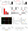
Figure 3. Far-Red Optogenetic Inhibition and Single-Component, Bidirectional Optical Control
(A) Six hundred thirty nanometer light evokes robust photocurrents in neurons transduced with eNpHR3.0 (representative voltage clamp trace at left). Summary plot comparing eNpHR2.0- and eNpHR3.0-expressing neurons (at right); eNpHR2.0, 42.7 ± 4.5 pA; eNpHR3.0, 239.4 ± 28.7 pA; unpaired t test p = 0.00004; n = 10). (B) Six hundred thirty nanometer illumination evoked robust hyperpolarization (representative voltage clamp trace at left). Summary plot comparing eNpHR2.0- and eNpHR3.0-expressing neurons (right); 15.6 ± 3.2 mV for eNpHR2.0 and 43.3 ± 6.1 mV for eNpHR3.0; unpaired t test p = 0.00116; n = 10). (C) Summary of outward photocurrents evoked by different wavelengths of red and far-red/infrared border illumination: 630 nm, 239.4 ± 28.7 pA (left, n = 10); 660 nm, 120.5 ± 16.7 pA (middle, n = 4); and 680 nm: 76.3 ± 9.1 pA (right, n = 4). Power density: 3.5 mW/mm2 (630 nm) and 7 mW/mm2 (660 nm, 680 nm). (D) Illumination with red and far-red/infrared border light inhibited spiking induced by current injection in neurons expressing eNpHR3.0. Typical current-clamp traces show optical inhibition at 630 nm (top left), 660 nm (top right), and 680 nm (bottom). Power density: 3.5 mW/mm2 (630 nm) and 7 mW/mm2 (660 nm, 680 nm). (E) eNpHR 3.0-EYFP (left) and hChR2-mCherry (middle) expressed both on the neuronal membrane and throughout the neurites. (F) Activation spectrum for eNPAC (left), and for ChR2(H134R) (right, blue) and eNpHR3.0 (right, yellow) alone. Maximum eNPAC steady-state excitation was 567 ± 49 pA at 427 nm (n = 9), 62% of the value for ChR2(H134R) alone (916 ± 185 pA; n = 5). Similarly, maximum eNPAC inhibition was 679 ± 109 pA at 590 nm (n = 9), 61% of the value for eNpHR3.0 alone (1110 ± 333 pA; n = 4). Output power density for peak eNpHR3.0 current values was 3.5–5 mW/mm2 (3.5 mW/mm2 at 590 nm). (G) Blue light (445 nm, 5 ms pulses) drove spiking at 20 Hz (left) and 10 Hz (right), while simultaneous application of yellow light (590 nm) inhibited spikes.
Figure 4
Figure 4. eBR: Trafficking-Enhanced Tool for Green Inhibition
(A) Confocal images in hippocampal neurons: unmodified BR forms aggregates similar to those of unmodified NpHR (top left). Provision of the TS motif before EYFP decreased aggregate size but did not fully eliminate aggregates (top right). Only when the ER export motif (FCYENEV) was also added to the C terminus were aggregates abolished, resulting in good membrane targeting throughout the soma (middle panels) and far into processes (bottom panels). (B) Five hundred sixty nanometer light (green bar) induced outward photocurrents in eBR cells (left, sample trace in voltage clamp), 46.4 ± 7.2 pA (right bar graph). Mean ± SEM is plotted, n = 12. Membrane input resistance was similar for all neurons patched (131.6 ± 19.5 mΩ). Light power density at sample was 7 mW/mm2. (C) Corresponding light-induced hyperpolarizations (left, sample trace in current clamp) were 10.8 ± 1.0 mV (right bar graph). Mean ± SEM is plotted, n = 12. (D) Illumination with green light (560 nm) sufficed to inhibit current injection-induced spiking. See also Figure S2.
Figure 5
Figure 5. Optogenetics: Molecular Design for Microbial Tools
(A) General subcellular targeting strategies for adapting microbial opsin genes to metazoan intact-systems biology. (B) Refinement of targeting at the tissue and subcellular levels. Subcellular opsin targeting methods have been previously described (for various dendritic compartment strategies, see Gradinaru et al. [2007] and Lewis et al., [2009]), and tissue/transcellular opsin targeting methods are described in Figure 2.

Comment in

  • Optogenetics 3.0.
    Liu X, Tonegawa S. Liu X, et al. Cell. 2010 Apr 2;141(1):22-4. doi: 10.1016/j.cell.2010.03.019. Cell. 2010. PMID: 20371341

References

    1. Adamantidis AR, Zhang F, Aravanis AM, Deisseroth K, de Lecea L. Neural substrates of awakening probed with optogenetic control of hypocretin neurons. Nature. 2007;450:420–424. - PMC - PubMed
    1. Airan RD, Thompson KR, Fenno LE, Bernstein H, Deisseroth K. Temporally precise in vivo control of intracellular signalling. Nature. 2009;458:1025–1029. - PubMed
    1. Aravanis AM, Wang LP, Zhang F, Meltzer LA, Mogri MZ, Schneider MB, Deisseroth K. An optical neural interface: in vivo control of rodent motor cortex with integrated fiberoptic and optogenetic technology. J. Neural Eng. 2007;4:S143–S156. - PubMed
    1. Arenkiel BR, Peca J, Davison IG, Feliciano C, Deisseroth K, Augustine GJ, Ehlers MD, Feng G. In vivo light-induced activation of neural circuitry in transgenic mice expressing channelrhodopsin-2. Neuron. 2007;54:205–218. - PMC - PubMed
    1. Arrenberg AB, Del Bene F, Baier H. Optical control of zebrafish behavior with halorhodopsin. Proc. Natl. Acad. Sci. USA. 2009;106:17968–17973. - PMC - PubMed

Publication types

Substances