Review
. 2010 Jun;31(17):4639-56.
doi: 10.1016/j.biomaterials.2010.02.044. Epub 2010 Mar 19.

Bioactive modification of poly(ethylene glycol) hydrogels for tissue engineering

Affiliations
Review

Bioactive modification of poly(ethylene glycol) hydrogels for tissue engineering

Junmin Zhu. Biomaterials. 2010 Jun.

Abstract

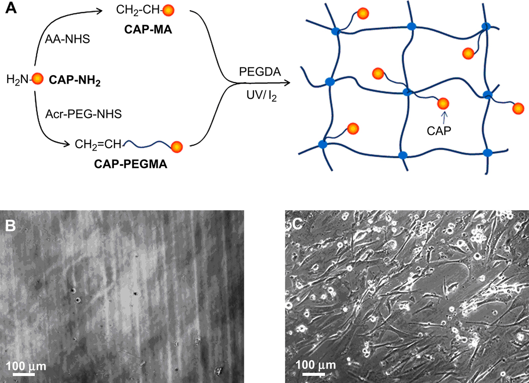
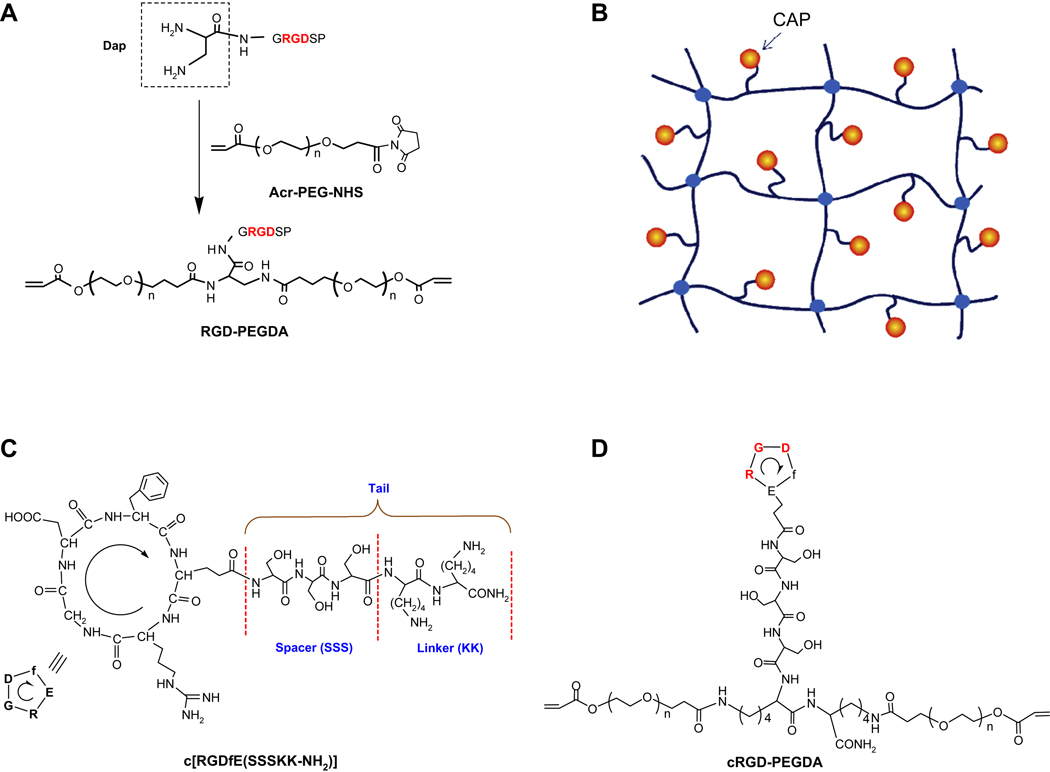
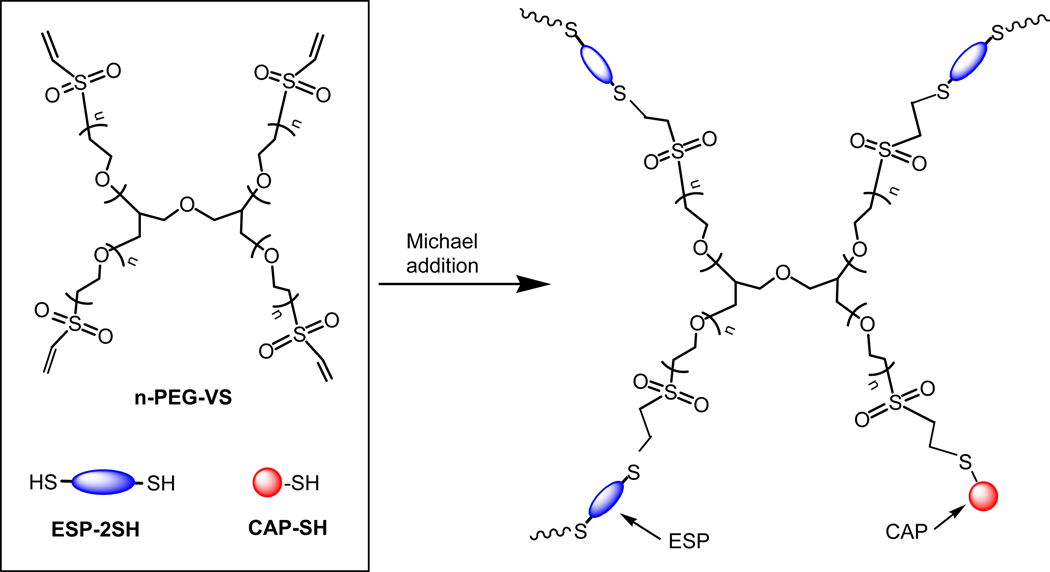
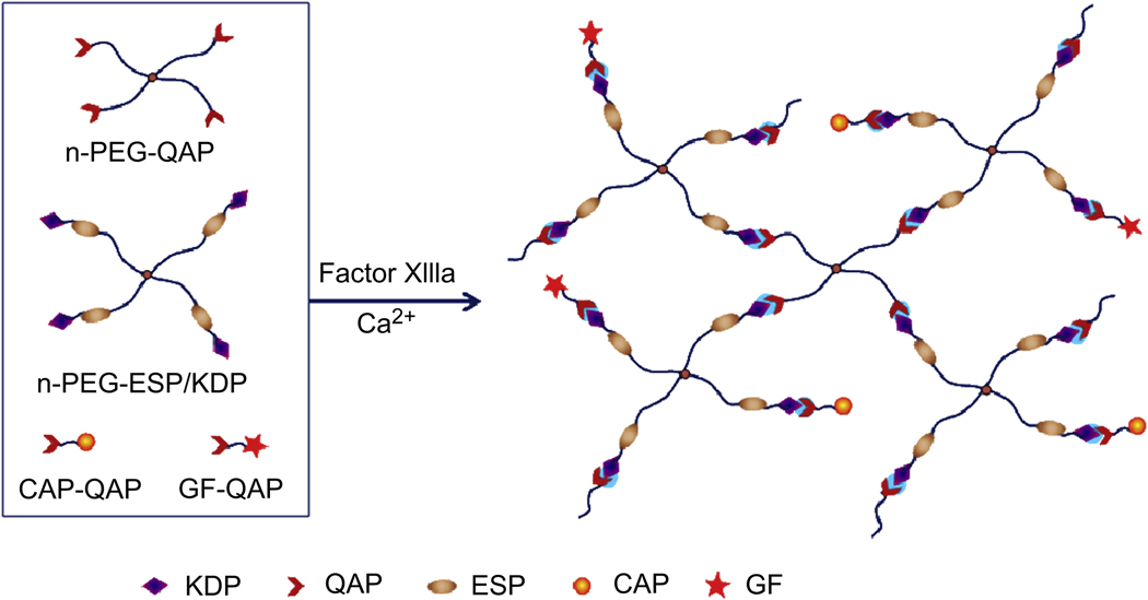
In this review, we explore different approaches for introducing bioactivity into poly(ethylene glycol) (PEG) hydrogels. Hydrogels are excellent scaffolding materials for repairing and regenerating a variety of tissues because they can provide a highly swollen three-dimensional (3D) environment similar to soft tissues. Synthetic hydrogels like PEG-based hydrogels have advantages over natural hydrogels, such as the ability for photopolymerization, adjustable mechanical properties, and easy control of scaffold architecture and chemical compositions. However, PEG hydrogels alone cannot provide an ideal environment to support cell adhesion and tissue formation due to their bio-inert nature. The natural extracellular matrix (ECM) has been an attractive model for the design and fabrication of bioactive scaffolds for tissue engineering. ECM-mimetic modification of PEG hydrogels has emerged as an important strategy to modulate specific cellular responses. To tether ECM-derived bioactive molecules (BMs) to PEG hydrogels, various strategies have been developed for the incorporation of key ECM biofunctions, such as specific cell adhesion, proteolytic degradation, and signal molecule-binding. A number of cell types have been immobilized on bioactive PEG hydrogels to provide fundamental knowledge of cell/scaffold interactions. This review addresses the recent progress in material designs and fabrication approaches leading to the development of bioactive hydrogels as tissue engineering scaffolds.


Figures

Fig. 1
Fig. 1
Structures of linear PEG and 4-arm-PEG with various functional end groups.
Fig. 2
Fig. 2
Model of complex 3D structure of extracellular matrix (ECM) and cell-ECM interactions.
Fig. 3
Fig. 3
Bioactive modification of PEG hydrogels (A) with bioactive molecules (BMs), such as cell-adhesive peptide (CAP), enzyme-sensitive peptide (ESP), and growth factor (GF), and major types of bioactive monomers from mono-, di- and multi-functionalization of BMs with various groups (B).
Fig. 4
Fig. 4
Fabrication of cell-adhesive PEG hydrogels (A) by copolymerization of PEGDA and acrylic acid, followed by post-grafting of cell-adhesive peptides (CAPs) on the hydrogel surface through the reaction between the N-terminal amino groups of CAPs and the carboxyl groups provided by acrylic acid from the hydrogel. Microfabrication of patterned cell-adhesive hydrogel surfaces (B) by microcontact printing (µCP) of biotinated CAPs (Biotin-CAPs) on streptavidin-bearing PEG hydrogels.
Fig. 5
Fig. 5
Synthesis of cell adhesive peptide (CAP) monoacrylamide (CAP-MA) and CAP-containing PEG monoacrylate (CAP-PEGMA) for preparation of cell-adhesive PEG hydrogels (A). Phase contrast images of human artery SMCs 6 h after seeding on 10% (w/v) PEGDA (Mw 6000) hydrogels (B) and on 10% (w/v) PEGDA (Mw 6000) hydrogels with incorporation of 0.5% (w/v) RGD-PEGMA (PEG Mw 3400; RGD sequence, GRGDSP, density ∼ 1 mM) (C).
Fig. 6
Fig. 6
Synthesis of RGD-containing PEGDA (RGD-PEGDA) (A) by conjugating diaminopropionic acid (Dap)-capped GRGDSP with Acr-PEG-NHS. Model of cell-adhesive PEG hydrogels (B) from photopolymerization of cell adhesive peptide (CAP)-containing PEGDA (CAP-PEGDA) with controlled spatial organization of CAPs. Structures of cyclic RGD (cRGD), c[(RGDfE)SSSKK(NH2)] (C) with a hydrophilic tail, consisting a spacer of three serine residues (SSS) and a linker of two lysine residues (KK), and cRGD-containing PEGDA (cRGD-PEGDA) (D).
Fig. 7
Fig. 7
Synthesis of enzyme-sensitive peptide (ESP)-containing PEGDA (ESP-PEGDA) by conjugating Acr-PEG-NHS with ESP diamine (ESP-2NH2), and preparation of proteolytically degradable PEG hydrogels from photopolymerization of ESP-PEGDA.
Fig. 8
Fig. 8
Preparation of cell-adhesive PEG hydrogels by thiol-acrylate photopolymerization of PEGDA with monothiol-containing cell adhesive peptide (CAP-SH).
Fig. 9
Fig. 9
Preparation of cell-adhesive PEG hydrogels by Michael addition of 4-arm-PEG vinyl sulfone (4-PEG-VS) with monothiol-containing cell-adhesive peptide (CAP-SH) and dithiol-containing enzyme-sensitive peptide (ESP-2SH).
Fig. 10
Fig. 10
Preparation of cell-adhesive PEG hydrogels by Click chemistry between 4-arm-PEG acetylene (4-PEG-Ace) and cell adhesive peptide diazide (CAP-2N3) in the presence of copper (II) sulfate and sodium ascorbate.
Fig. 11
Fig. 11
Preparation of bioactive PEG hydrogels by enzymatic reaction between Lys donor peptide (KDP)-capped multiarm PEG (n-PEG-KDP) and Gln acceptor peptide (QAP)-capped enzyme-sensitive peptide (ESP)-containing multiarm PEG (n-PEG-ESP/QAP) with incorporation of QAP-functionalized cell adhesive peptide (CAP-QAP) and/or QAP-functionalized growth factor (GF-QAP) in the presence of Factor XIIIa and calcium ion.
Fig. 12
Fig. 12
Photodegradable peptide backbone (A) containing the residue of 2-(nitrophenyl)glycine (NPG), and photoisomerizable peptide backbone (B) containing the residue of 4-[(4-amino)phenylazo]benzoic acid (APB).
Fig. 13
Fig. 13
Preparation of photocleavable cell-adhesive PEG hydrogels by copolymerization of PEGDA with photocleavable cell-adhesive peptide monoacrylate (PD-CAPMA), and photodegradable PEG hydrogels by copolymerization of photocleaveable PEGDA (PD-PEGDA) with PEG monoacrylate (PEGMA).

References

    1. Langer R, Vacanti JP. Tissue engineering. Science. 1993;260:920–926. - PubMed
    1. Hoffman AS. Hydrogels for biomedical applications. Adv Drug Deliver Rev. 2002;43:3–12. - PubMed
    1. Lee KY, Mooney DJ. Hydrogels for tissue engineering. Chem Rev. 2001;101:1869–1879. - PubMed
    1. Temenoff JS, Mikos AG. Injectable biodegradable materials for orthopedic tissue engineering. Biomaterials. 2002;21:2405–2412. - PubMed
    1. Elisseeff J, Anseth K, Sims D, McIntosh W, Randolph M, Langer R. Transdermal photopolymerization for minimally invasive implantation. Proc Natl Acad Sci U S A. 1999;96:3104–3107. - PMC - PubMed

Publication types

MeSH terms