

Correction of a carboxyl terminal simian immunodeficiency virus Nef frameshift mutation restores virus replication in macaques
- PMID: 20303562
- PMCID: PMC3418331
- DOI: 10.1016/j.virol.2010.02.026
Correction of a carboxyl terminal simian immunodeficiency virus Nef frameshift mutation restores virus replication in macaques
Abstract
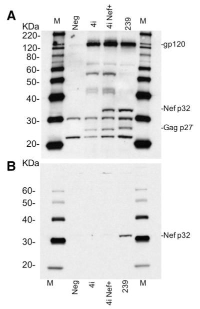
Previous studies demonstrated that the nef gene is a critical determinant of the pathogenicity of simian immunodeficiency virus (SIV) in macaques. In the present study, we evaluated the effect of a spontaneous frameshift mutation in the C-terminus of the nef gene of the minimally pathogenic SIVsmH4i clone. This clone exhibited a single nucleotide deletion in the nef gene relative to pathogenic SIV clones that resulted in a frameshift and addition of 46 amino acids to the C-terminus of Nef. We generated a corrected version of this clone, SIVsmH4i Nef+ that restored Nef protein expression. Inoculation of macaques with SIVsmH4i resulted in delayed and low levels of peak viremia. This contrasted with improved kinetics and robust peak viremia in macaques inoculated with the corrected version. Despite the restoration of in vivo replication ability, neither clone resulted in memory CD4+ T cell loss or disease in a period of two years.
Published by Elsevier Inc.
Figures


Similar articles
-
Chronology of genetic changes in the vpu, env, and Nef genes of chimeric simian-human immunodeficiency virus (strain HXB2) during acquisition of virulence for pig-tailed macaques.Virology. 1998 Sep 1;248(2):275-83. doi: 10.1006/viro.1998.9300. Virology. 1998. PMID: 9721236
-
The ITAM in Nef influences acute pathogenesis of AIDS-inducing simian immunodeficiency viruses SIVsm and SIVagm without altering kinetics or extent of viremia.J Virol. 2002 May;76(9):4379-89. doi: 10.1128/jvi.76.9.4379-4389.2002. J Virol. 2002. PMID: 11932405 Free PMC article.
-
A truncated form of Nef selected during pathogenic reversion of simian immunodeficiency virus SIVmac239Deltanef increases viral replication.J Virol. 2003 Jan;77(2):1245-56. doi: 10.1128/jvi.77.2.1245-1256.2003. J Virol. 2003. PMID: 12502842 Free PMC article.
-
Immunodeficiency viruses. Not enough sans Nef.Curr Biol. 1994 Jul 1;4(7):618-20. doi: 10.1016/s0960-9822(00)00135-4. Curr Biol. 1994. PMID: 7953537 Review.
-
Nef and PAK: virulence factor and cellular accomplice.Chem Biol. 1997 Jan;4(1):13-5. doi: 10.1016/s1074-5521(97)90232-5. Chem Biol. 1997. PMID: 9070423 Review.
References
-
- Arien KK, Verhasselt B. HIV Nef: role in pathogenesis and viral fitness. Curr. HIV Res. 2008;6(3):200–208. - PubMed
-
- Arold ST, Baur AS. Dynamic Nef and Nef dynamics: how structure could explain the complex activities of this small HIV protein. Trends Biochem. Sci. 2001;26(6):356–363. - PubMed
-
- Atkins KM, Thomas L, Youker RT, Harriff MJ, Pissani F, You H, Thomas G. HIV-1 Nef binds PACS-2 to assemble a multikinase cascade that triggers major histocompatibility complex class I (MHC-I) down-regulation: analysis using short interfering RNA and knock-out mice. J. Biol. Chem. 2008;283(17):11772–11784. - PMC - PubMed
-
- Baba TW, Liska V, Hofmann-Lehmann R, Vlasak J, Xu W, Ayehunie S, Cavacini LA, Posner MR, Katinger H, Stiegler G, Bernacky BJ, Rizvi TA, Schmidt R, Hill LR, Keeling ME, Lu Y, Wright JE, Chou TC, Ruprecht RM. Human neutralizing monoclonal antibodies of the IgG1 subtype protect against mucosal simian-human immunodeficiency virus infection. Nat. Med. 2000;6(2):200–206. - PubMed
Publication types
MeSH terms
Substances
Grants and funding
LinkOut - more resources
Full Text Sources
Research Materials
