

Effect of genetic deletion of the vanilloid receptor TRPV1 on the expression of Substance P in sensory neurons of mice with adjuvant-induced arthritis
- PMID: 20303589
- PMCID: PMC2879442
- DOI: 10.1016/j.npep.2010.02.003
Effect of genetic deletion of the vanilloid receptor TRPV1 on the expression of Substance P in sensory neurons of mice with adjuvant-induced arthritis
Abstract
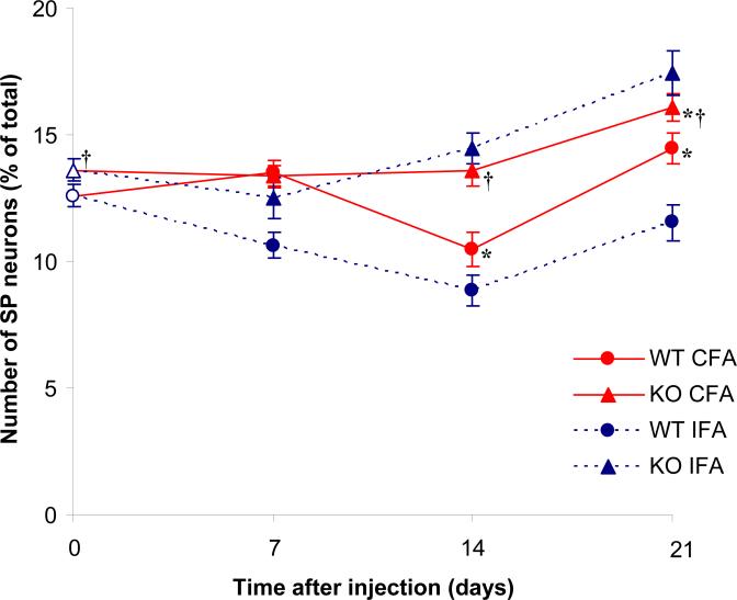
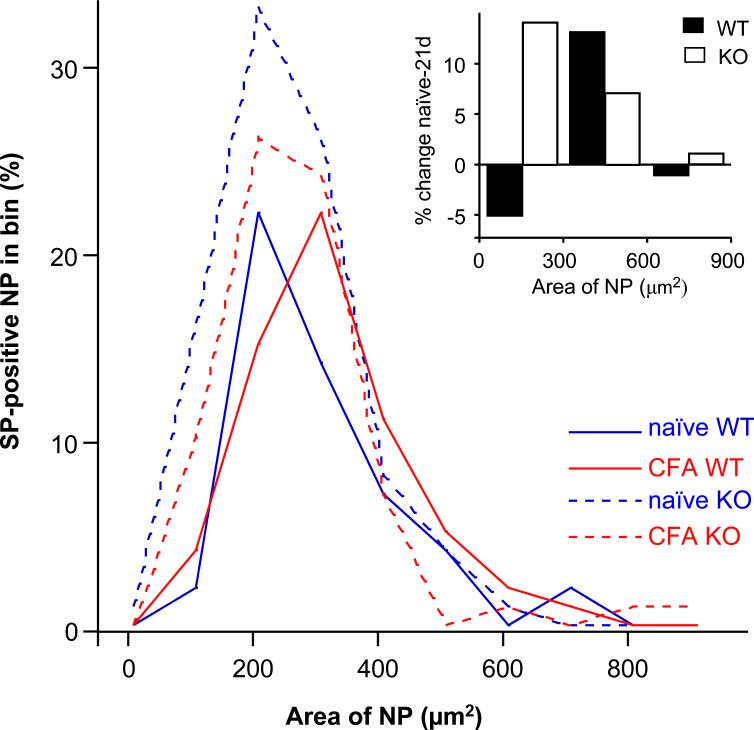
The neuropeptide Substance P (SP), expressed by nociceptive sensory afferents in joints, plays an important role in the pathogenesis of arthritis. Capsaicin causes neurons in the dorsal root ganglia (DRG) to release SP from their central and peripheral axons, suggesting a functional link between SP and the capsaicin receptor, the transient receptor potential vanilloid 1 (TRPV1). The expression of both TRPV1 and SP have been reported to increase in several models of arthritis but the specific involvement of TRPV1-expressing articular afferents that can release SP is not completely understood. We here wanted to ascertain whether the increase in the number of SP-positive primary afferents in arthritis may be affected by genetic deletion of TRPV1. For this, we used immunohistochemistry to quantify the expression of SP in primary afferent neurons in wild-type mice (WT) vs. TRPV1-knockout (KO) mice with adjuvant-induced arthritis (AIA). We found that the expression of SP in DRG (1) increased significantly over naïve level in both WT and KO mice 3 weeks after AIA, (2) was significantly higher in KO mice than in WT mice in naïve mice and 2-3 weeks after AIA, (3) was significantly higher on the side of AIA than on the contralateral, vehicle-injected side at all time points in WT mice, but not in KO mice, and (4) increased predominantly in small-size neurons in KO mice and in small- and medium-size neurons in WT mice. Since the size distribution of SP-positive DRG neurons in arthritic TRPV1-KO mice was not significantly different from that in naïve mice, we speculate that the increased expression of SP is unlikely to reflect recruitment of A-fiber primary afferents and that the higher expression of SP in KO mice may represent a plastic change to compensate for the missing receptor in a major sensory circuit.
Copyright 2010 Elsevier Ltd. All rights reserved.
Figures
Similar articles
-
Increased expression of CGRP in sensory afferents of arthritic mice--effect of genetic deletion of the vanilloid receptor TRPV1.Neuropeptides. 2008 Oct-Dec;42(5-6):551-6. doi: 10.1016/j.npep.2008.08.001. Epub 2008 Sep 11. Neuropeptides. 2008. PMID: 18789524 Free PMC article.
-
Vanilloid receptor TRPV1-mediated phosphorylation of ERK in murine adjuvant arthritis.Osteoarthritis Cartilage. 2009 Feb;17(2):244-51. doi: 10.1016/j.joca.2008.06.015. Epub 2008 Aug 5. Osteoarthritis Cartilage. 2009. PMID: 18684647 Free PMC article.
-
Influence of the vanilloid receptor TRPV1 on the activation of spinal cord glia in mouse models of pain.Exp Neurol. 2009 Dec;220(2):383-90. doi: 10.1016/j.expneurol.2009.09.030. Epub 2009 Oct 6. Exp Neurol. 2009. PMID: 19815011 Free PMC article.
-
Vanilloid receptor TRPV1-positive sensory afferents in the mouse ankle and knee joints.Brain Res. 2008 Jul 11;1219:59-65. doi: 10.1016/j.brainres.2008.04.043. Epub 2008 Apr 27. Brain Res. 2008. PMID: 18538749 Free PMC article.
-
The role of Transient Receptor Potential Vanilloid 1 (TRPV1) channels in pancreatitis.Biochim Biophys Acta. 2007 Aug;1772(8):869-78. doi: 10.1016/j.bbadis.2007.02.012. Epub 2007 Mar 12. Biochim Biophys Acta. 2007. PMID: 17428642 Free PMC article. Review.
Cited by
-
Mechanisms Underlying the Scratching Behavior Induced by the Activation of Proteinase-Activated Receptor-4 in Mice.J Invest Dermatol. 2015 Oct;135(10):2484-2491. doi: 10.1038/jid.2015.183. Epub 2015 May 8. J Invest Dermatol. 2015. PMID: 25955385
-
Skin reaction to capsaicin in patients with systemic lupus erythematosus compared to healthy controls.Caspian J Intern Med. 2021 Mar;12(2):140-147. doi: 10.22088/cjim.12.2.140. Caspian J Intern Med. 2021. PMID: 34012530 Free PMC article.
-
Hemokinin-1 as a Mediator of Arthritis-Related Pain via Direct Activation of Primary Sensory Neurons.Front Pharmacol. 2021 Jan 13;11:594479. doi: 10.3389/fphar.2020.594479. eCollection 2020. Front Pharmacol. 2021. PMID: 33519457 Free PMC article.
-
Long term follow-up of heart rate variability in healthcare workers with mild COVID-19.Front Neurol. 2024 May 17;15:1403551. doi: 10.3389/fneur.2024.1403551. eCollection 2024. Front Neurol. 2024. PMID: 38827576 Free PMC article.
-
Role of capsaicin-sensitive nerves and tachykinins in mast cell tryptase-induced inflammation of murine knees.Inflamm Res. 2016 Sep;65(9):725-36. doi: 10.1007/s00011-016-0954-x. Epub 2016 Jun 1. Inflamm Res. 2016. PMID: 27251170
References
-
- Abramovici A, Daizade I, Yosipovitch Z, Gibson SJ, Polak JM. The distribution of peptide-containing nerves in the synovia of the cat knee joint. Histol Histopathol. 1991;6:469–476. - PubMed
-
- Ahmed M, Bjurholm A, Schultzberg M, Theodorsson E, Kreicbergs A. Increased levels of substance P and calcitonin gene-related peptide in rat adjuvant arthritis. A combined immunohistochemical and radioimmunoassay analysis. Arthritis Rheum. 1995a;38:699–709. - PubMed
-
- Ahmed M, Bjurholm A, Srinivasan GR, Lundeberg T, Theodorsson E, Schultzberg M, Kreicbergs A. Capsaicin effects on substance P and CGRP in rat adjuvant arthritis. Regul Pept. 1995b;55:85–102. - PubMed
-
- Bar KJ, Schaible HG, Brauer R, Halbhuber KJ, von Banchet GS. The proportion of TRPV1 protein-positive lumbar DRG neurones does not increase in the course of acute and chronic antigen-induced arthritis in the knee joint of the rat. Neurosci Lett. 2004;361:172–175. - PubMed
-
- Barton NJ, McQueen DS, Thomson D, Gauldie SD, Wilson AW, Salter DM, Chessell IP. Attenuation of experimental arthritis in TRPV1R knockout mice. Exp Mol Pathol. 2006;81:166–170. - PubMed
Publication types
MeSH terms
Substances
Grants and funding
LinkOut - more resources
Full Text Sources
Research Materials
