. 2010 Aug;35(1):45-51.
doi: 10.1016/j.jaut.2010.01.004. Epub 2010 Mar 19.

Tg.2098 is a major human thyroglobulin T-cell epitope

Affiliations

Tg.2098 is a major human thyroglobulin T-cell epitope

Francesca Menconi et al. J Autoimmun. 2010 Aug.

Abstract

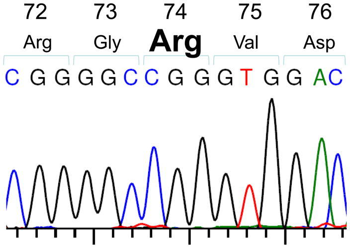
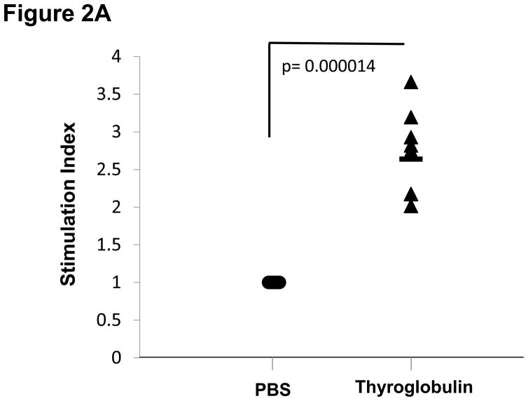
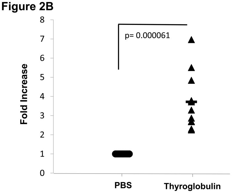
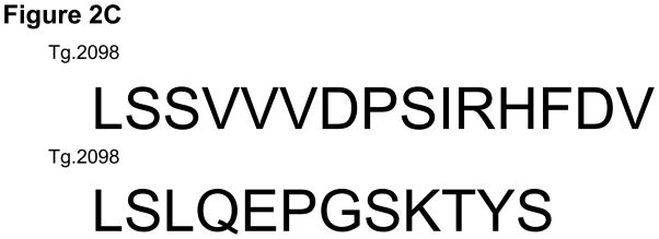
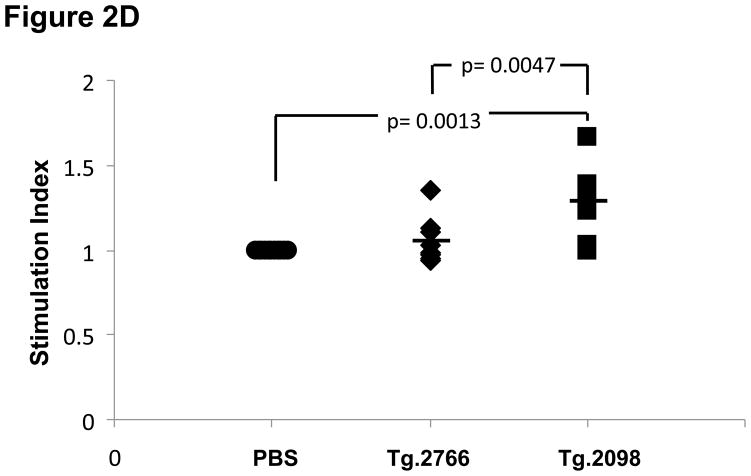
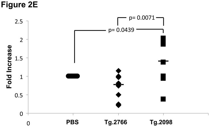
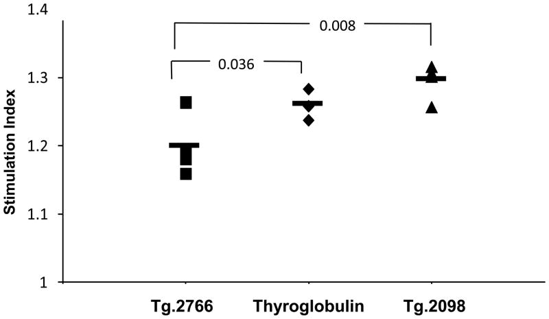
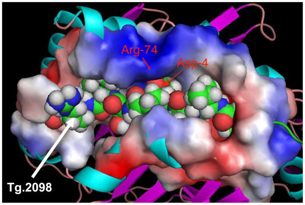
An HLA-DR variant containing Arginine at position 74 of the DRbeta1 chain confers a strong genetic susceptibility to autoimmune thyroid diseases (AITD), Graves' disease (GD) and Hashimoto's thyroiditis (HT), while Glutamine at position DRbeta1-74 is protective. We hypothesized that the DRbeta1-Arg74 variant is able to present pathogenic thyroglobulin (Tg) peptides to T-cells more efficiently, thereby triggering thyroid autoimmunity. Indeed, we have previously identified 4 human Tg (hTg) peptides that bind specifically to DRbeta1-Arg74 with much weaker binding to the protective variant DRbeta1-Gln74. The aim of our study was to examine in vivo whether an hTg peptide that binds strongly and specifically to DRbeta1-Arg74 is capable of stimulating T-cells during the induction of thyroiditis in a "humanized" mouse expressing human DR3, and in patients positive for Tg antibodies. Sequencing of exon 2 of the DR transgene in the DR3 mice, null for endogenous MHC II molecules, confirmed that they expressed the disease-associated DRbeta1-Arg74 variant, thus making them an ideal in vivo model to test the presentation of hTg peptides by DRbeta1-Arg74 HLA-DR. Induction of EAT in the DR3 mice lead to T-cell stimulation and proliferation to Tg.2098, a strong and specific DRbeta1-Arg74 binder, while a non-binding control peptide, Tg.2766 did not induce this response. Moreover, Tg.2098 stimulated T-cells from 4 individuals who were positive for thyroglobulin antibodies, demonstrating that Tg.2098 is an immunogenic peptide capable of being presented in vivo and activating T-cells in EAT and AITD. Energetic analysis of the complex formed by Tg.2098 and DRbeta-Arg74 has shown that the origin of the affinity was determined by residues 1, 7 and 9 in the peptide, while the selectivity of the peptide for the MHC was determined by the Asp in position 4. The disease-protective substitution R74Q, leads to reduction in affinity due to changes in local interaction with D4 as well as non-local interaction with other residues. The electrostatic potential on the surface of the DRbeta-Arg74-Tg.2098 complex has a unique signature which may be recognized by T-cell receptors leading to autoimmune thyroiditis. Taken together these findings suggest that Tg.2098, a strong and specific binder to the disease-associated HLA-DRbeta-Arg74, is a major human T-cell epitope and participant in the pathoetiology of AITD.


Figures

Figure 1
Figure 1
Sequencing of exon 2 of the DRβ1 gene in DR3 transgenic mice demonstrates the presence of arginine (CGG) at position 74 of the DRβ1 chain.
Figure 2
Figure 2
EAT was induced in DR3 mice by injection of full length human thyroglobulin with complete Freund’s adjuvant. After 28 days the mice were sacrificed and T-cells were collected from draining lymph nodes (inguinal and cervical) and spleen. (A) Lymphocytes from mice immunized with hTg were incubated with hTg and proliferation was measured by 3[H]-Thymidine incorporation. Significant proliferation is seen in lymphocytes incubated with hTg compared to PBS. (B) Levels of IL-2 production by lymphocytes incubated with hTG were also significantly elevated. (C) Sequences of the Tg.2098 and Tg.2766 peptide tested in cellular proliferation assays, sequences showed no significant sequence alignment. (D) Lymphocytes from mice immunized with hTg were incubated with hTg peptides Tg.2098 and Tg.2766 and proliferation was measured by 3[H]-Thymidine incorporation. Significant proliferation is seen in lymphocytes incubated with Tg.2098 compared to lymphocytes incubated with PBS or with the non-binding peptide Tg.2766. (E) Levels of IL-2 production by lymphocytes incubated with Tg.2098 were significantly elevated compared to lymphocytes incubated with PBS or with the non-binding peptide Tg.2766.
Figure 2
Figure 2
EAT was induced in DR3 mice by injection of full length human thyroglobulin with complete Freund’s adjuvant. After 28 days the mice were sacrificed and T-cells were collected from draining lymph nodes (inguinal and cervical) and spleen. (A) Lymphocytes from mice immunized with hTg were incubated with hTg and proliferation was measured by 3[H]-Thymidine incorporation. Significant proliferation is seen in lymphocytes incubated with hTg compared to PBS. (B) Levels of IL-2 production by lymphocytes incubated with hTG were also significantly elevated. (C) Sequences of the Tg.2098 and Tg.2766 peptide tested in cellular proliferation assays, sequences showed no significant sequence alignment. (D) Lymphocytes from mice immunized with hTg were incubated with hTg peptides Tg.2098 and Tg.2766 and proliferation was measured by 3[H]-Thymidine incorporation. Significant proliferation is seen in lymphocytes incubated with Tg.2098 compared to lymphocytes incubated with PBS or with the non-binding peptide Tg.2766. (E) Levels of IL-2 production by lymphocytes incubated with Tg.2098 were significantly elevated compared to lymphocytes incubated with PBS or with the non-binding peptide Tg.2766.
Figure 3
Figure 3
Lymphocytes from 4 patients who are positive for anti-Tg antibodies were incubated with hTg, and hTg peptides Tg.2098 and Tg.2766. Proliferation was measured by 3[H]-Thymidine incorporation. Significant proliferation is seen in lymphocytes incubated with Tg.2098 compared to lymphocytes incubated with the non-binding peptide Tg.2766.
Figure 4
Figure 4
Tg.2098 embedded inside the binding groove, with a characteristic and unique electrostatic potentials displayed on its surface. The ridge opposite the Arg-74 shows a positive potential flanked by negative potential.

Similar articles

Cited by

References

    1. Castellino F, Zhong G, Germain RN. Antigen presentation by MHC class II molecules: invariant chain function, protein trafficking, and the molecular basis of diverse determinant capture. Hum Immunol. 1997;54(2):159–169. - PubMed
    1. Jacobson EM, Huber A, Tomer Y. The HLA gene complex in thyroid autoimmunity: From epidemiology to etiology. J Autoimmun. 2008;30(1–2):58–62. - PMC - PubMed
    1. Lee KH, Wucherpfennig KW, Wiley DC. Structure of a human insulin peptide-HLA-DQ8 complex and susceptibility to type 1 diabetes. Nat Immunol. 2001;2(6):501–507. - PubMed
    1. Wucherpfennig KW. MHC-linked susceptibility to type 1 diabetes: a structural perspective. Ann N Y Acad Sci. 2003;1005:119–127. - PubMed
    1. Onengut-Gumuscu S, Concannon P. The genetics of type 1 diabetes: lessons learned and future challenges. J Autoimmun. 2005;25(Suppl):34–39. - PubMed

Publication types

MeSH terms