Review
. 2010 Jun;20(6):363-9.
doi: 10.1016/j.tcb.2010.02.007. Epub 2010 Mar 19.

p53 and ARF: unexpected players in autophagy

Affiliations
Review

p53 and ARF: unexpected players in autophagy

Gregor M Balaburski et al. Trends Cell Biol. 2010 Jun.

Abstract

p53 and ARF are well-established tumor-suppressor proteins that function together in the negative regulation of cancer. Recently, both proteins were found to play surprising roles in autophagy. Autophagy ('self-eating') is a crucial response of eukaryotic cells to metabolic and other stress. During this process, portions of the cytosol are sequestered into characteristic double-membrane vesicles that are delivered to the lysosome for degradation, leading to the release of free amino acids and promoting cell survival. The mechanisms whereby p53 and ARF control autophagy are only now becoming elucidated. An emerging question is whether we can develop metabolic poisons that preferentially destroy tumor cells depending on their reliance on autophagy for survival, and on their p53 and ARF status.


Figures

Figure 1
Figure 1. The p53-ARF axis in cancer and autophagy
A) In the p53-ARF-MDM2 pathway in cancer, oncogenes transcriptionally induce ARF, which binds and inhibits MDM2, leading to stabilization of p53. In two feedback loops, p53 positively regulates MDM2 transcription, and negatively regulates ARF transcription. B) In the p53-ARF axis in autophagy, p53 and ARF positively regulate autophagy in stressed cells. In contrast, in the unstressed cell, p53 negatively regulates ARF via transcriptional repression, and it also negatively regulates autophagy.
Figure 2
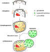
Figure 2. Major cellular events during autophagy
i) Nucleation: appearance of double layered, isolation membrane, the phagophore, ii) Elongation: extension of the phagophore, iii) autophagosome formation, encapsulation of cellular components and iv) Maturation: fusion of the autophagosome with the lysosome, resulting in formation of the autophagolysosome. Ultimately, the components in the autophagolysosome are degraded by lysosomal hydrolyses and the breakdown products are recycled in cellular bio-synthetic pathways.
Figure 3
Figure 3. Pathways upstream and downstream from p53-mediated autophagy
p53 is downstream of both nutrient stress and genotoxic stress. In nutrient-deprived cells, such as following glucose deprivation, p53 is phosphorylated and induces G1 arrest, thereby allowing for cell survival. In response to genotoxic stress, p53 likewise becomes active as a transcription factor, and transactivates a number of genes with direct roles in autophagy, such as DRAM, or that encode inhibitors of mTOR activity, including Sestrin 1 and 2, TSC2 and the β1 and -2 subunits of AMPK. In the unstressed cell, silencing or inhibiting p53 induces autophagy. Therefore, p53 is intimately tied to autophagy as both a positive and negative regulator, depending on the presence of stress.
Figure 4
Figure 4. Regulation of autophagy by p53 and ARF
Based on cellular localization and type of stress p53 can be a positive or negative regulator of autophagy. In the nucleus p53 transcriptionally regulates a number of autophagy modulators. Cytosolic p53 inhibits autophagy in unstressed cells. ARF positively regulates autophagy by disrupting the Bcl-xl/Beclin 1 complex, releasing Beclin-1 to induce autophagy. A truncated form of ARF, smARF directly localizes to the mitochondria to induce autophagy.

Similar articles

Cited by

References

    1. Vousden KH, Prives C. Blinded by the Light: The Growing Complexity of p53. Cell. 2009;137:413–431. - PubMed
    1. Eischen CM, et al. Disruption of the ARF-Mdm2-p53 tumor suppressor pathway in Myc-induced lymphomagenesis. Genes Dev. 1999;13:2658–2669. - PMC - PubMed
    1. Robertson KD, Jones PA. The human ARF cell cycle regulatory gene promoter is a CpG island which can be silenced by DNA methylation and down-regulated by wild-type p53. Mol Cell Biol. 1998;18:6457–6473. - PMC - PubMed
    1. Stott FJ, et al. The alternative product from the human CDKN2A locus, p14(ARF), participates in a regulatory feedback loop with p53 and MDM2. EMBO J. 1998;17:5001–5014. - PMC - PubMed
    1. Esclatine A, et al. Macroautophagy signaling and regulation. Curr Top Microbiol Immunol. 2009;335:33–70. - PubMed

Publication types

MeSH terms

Substances