Review
. 2010 Apr;16(4):193-202.
doi: 10.1016/j.molmed.2010.02.003. Epub 2010 Mar 19.

Regenerative medicine for retinal diseases: activating endogenous repair mechanisms

Affiliations
Review

Regenerative medicine for retinal diseases: activating endogenous repair mechanisms

M O Karl et al. Trends Mol Med. 2010 Apr.

Abstract

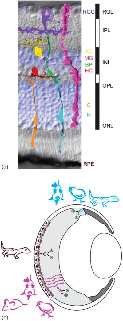
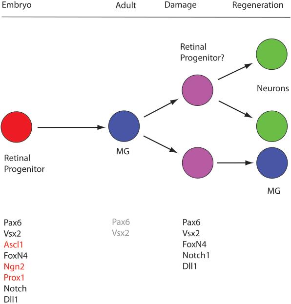
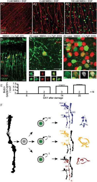
The retina is subject to degenerative diseases that often lead to significant visual impairment. Non-mammalian vertebrates have the remarkable ability to replace neurons lost through damage. Fish, and to a limited extent birds, replace lost neurons by the dedifferentiation of Müller glia to a progenitor state followed by the replication of these neuronal progenitor cells. Over the past five years, studies have investigated whether regeneration can be stimulated in the mouse and rat retina. Several groups have reported that at least some types of neurons can be regenerated in the mammalian retina in vivo or in vitro, and that the regeneration of neurons can be stimulated using growth factors, transcription factors or subtoxic levels of excitatory amino acids. These recent results suggest that some part of the regenerative program that occurs in non-mammalian vertebrates remains in the mammalian retina, and could provide a basis to develop new strategies for retinal repair in patients with retinal degenerations.


Figures

Figure 1
Figure 1
(a) Vertebrate retinas share a common architecture. 7 major cell types, 5 neuronal and 2 supporting, are regularly spaced across the retina. The background of the figure is an image (Nomarski contrast) of a mouse retinal cross section counterstained with a nuclear dye (DAPI in blue) showing the laminar structure: the inner and outer nuclear layer (INL and ONL) as well as the retinal ganglion cell layer (RGL) contain most of the cells (blue). The inner and outer plexiform layer (IPL and OPL) are made up of the axons and dendrites of the neurons. Light first crosses all layers to be detected by the light sensitive photoreceptor cells, rods (R, low light level sensors) and cones (C, daylight color sensors). If cones detect a change in light intensity they signal through different bipolar cells subtypes (BC) to the retinal ganglion cells (RGC), which send their axons in the optic nerve to the higher visual centers in the brain. Rod signals are relayed via rod bipolars, amacrine (AC) neurons, and/or cone bipolars to the ganglion cells. The rod and cone pathways are modified by inhibitory neurons, at the photoreceptor-bipolar synapse by horizontal cells (HC) and at the bipolar-ganglion synapse by amacrines. The retina also contains 3 major types of support cells: 1. the retinal pigment epithelium (RPE), 2. the Müller glia (MG) and 3. The astrocytes (not shown). (b) Depending on the species retinas may regenerate from two major cell sources, RPE and Müller glia, and may even have an active stem cell zone, the ciliary marginal zone (CMZ), analogous to stem cell zones like the subventricular zone of the brain. Progenitor cells (blue) in amphibian and fish CMZ generate most of the mature retina, but only a small part in birds. The CMZ can persist throughout the animal’s life and respond to injury with increased neurogenesis. In mammals no significant persistent neurogenic CMZ has been found. Upon damage of the central retina in amphibians, the RPE cells (brown) de-differentiate, proliferate and regenerate neurons; in fish and to a limited extent in birds and rodents, retinal damage causes Müller glia (magenta) to de-differentiate, proliferate and regenerate neurons (see Figure 3).
Figure 2
Figure 2
Progenitor genes are re-expressed in the damaged retina. The Muller glia are generated during embryonic development from the retinal progenitors, the same cells that produce all the other retinal cell types described in Figure 1. In the mature retina of mice, the Muller glia are mitotically quiescent and no longer express most of the progenitor genes; however, some of the progenitor genes are up-regulated after damage and when the cells are induced to re-enter the cell cycle by treating the retina with growth factors/mitogens. This suggests that at least some of the Muller cells in the mature mammalian retina can de-differentiate to a partial progenitor state, and it may explain why the cells are able to regenerate a subset of retinal neurons.
Figure 3
Figure 3
Retinal regeneration in mice. A) In vivo, intraocular application of neurotoxin NMDA leads to loss of retinal ganglion cells (indicated by neurofilament stain; red). Müller glia re-enter cell cycle after neuronal cell loss when treated with a mitogen, like EGF (single application; A2, A3), but not so much with either treatment alone (A1: EGF only, NMDA only not shown but similar to A1). BrdU was applied starting on day 2 after damage (D2) to identify proliferation. B) After NMDA damage GFAP-Cre::RYFP labels Müller glia which upregulate PCNA after daily injections of FGF1 (48 hours after NMDA, D2) for four subsequent days (analyzed on D6). Interestingly, in fish and chick, damage is sufficient to stimulate Müller glia proliferation and multiple cell divisions lead to a larger number of progeny (see summary Fig. 3F). C) After damage and growth factor treatment (FGF1 plus insulin) of mouse retina in vivo, BrdU-positive cells start to express Pax6 (not shown) and GAD67-GFP 8 to 30 days later. GAD67-GFP mice express GFP in immature neurons, a subset of mature RGCs, horizontal and all GABAergic amacrine cells in adult mouse retina. BrdU and GAD67-GFP double-labeled cells are found, like amacrine neurons, in the inner nuclear (BrdU+ Prox1+ GAD67-GFP+) and retinal ganglion cell layer (not shown). D) At D30, some BrdU-positive GAD67-GFP+ labeled cells also express Prox1 (boxed cell) suggesting amacrine cell regeneration. E) Graph shows the number of BrdU-positive /GAD67-GFP-positive cells as a function of days after NMDA damage. F) In comparison with fish and chick, retinal regeneration in mice is limited not only due to a restricted number of Müller glia re-entering the cell cycle and generation of an insufficient number of progeny cells, but most interestingly in the limited number and types of neurons regenerated (colored /labeled cells are regenerated in the respective species; RGC, retinal ganglion cell; AC, amacrine neuron; BP, bipolar neuron; HC, horizontal neuron; R, rod- and C, cone-photoreceptor; MG, Müller glia). Images show z-axis projection of (c) 2 × 1μm and (d) 1 × 1μm. Scale bars: 5 μm in orthogonal and inset views; 10 μm all other views. (Figures A-E from [39]).

References

    1. Jones BW, et al. Retinal remodeling triggered by photoreceptor degenerations. J Comp Neurol. 2003;464:1–16. - PubMed
    1. Fariss RN, et al. Abnormalities in rod photoreceptors, amacrine cells, and horizontal cells in human retinas with retinitis pigmentosa. Am J Ophthalmol. 2000;129:215–223. - PubMed
    1. Moshiri A, et al. Retinal stem cells and regeneration. Int J Dev Biol. 2004;48:1003–1014. - PubMed
    1. Tropepe V, et al. Retinal stem cells in the adult mammalian eye. Science. 2000;287:2032–2036. - PubMed
    1. Cicero SA, et al. Cells previously identified as retinal stem cells are pigmented ciliary epithelial cells. Proc Natl Acad Sci U S A. 2009;106:6685–6690. - PMC - PubMed