Review
. 2010 Mar 19;140(6):918-34.
doi: 10.1016/j.cell.2010.02.016.

Mechanisms underlying inflammation in neurodegeneration

Affiliations
Review

Mechanisms underlying inflammation in neurodegeneration

Christopher K Glass et al. Cell. .

Abstract

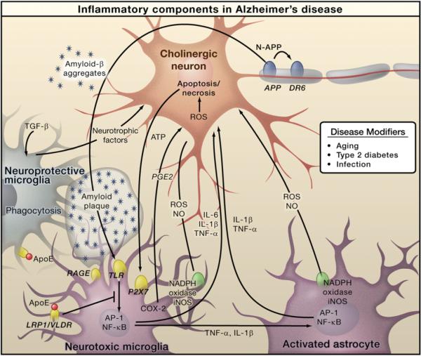
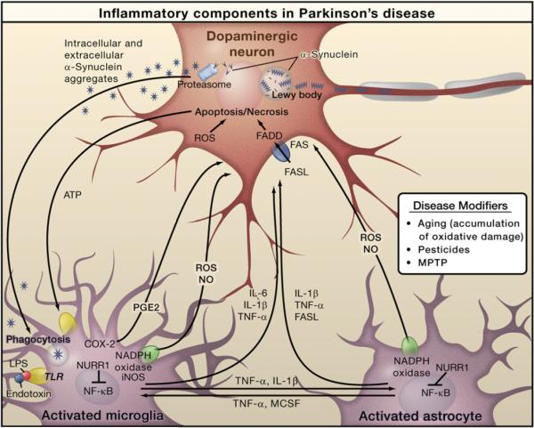
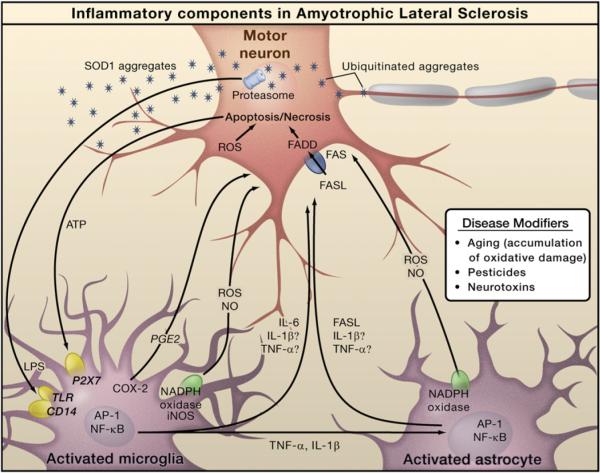
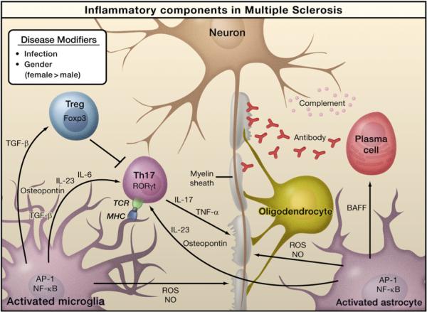
Inflammation is associated with many neurodegenerative diseases, including Alzheimer's disease, Parkinson's disease, amyotrophic lateral sclerosis, and multiple sclerosis. In this Review, we discuss inducers, sensors, transducers, and effectors of neuroinflammation that contribute to neuronal dysfunction and death. Although inducers of inflammation may be generated in a disease-specific manner, there is evidence for a remarkable convergence in the mechanisms responsible for the sensing, transduction, and amplification of inflammatory processes that result in the production of neurotoxic mediators. A major unanswered question is whether pharmacological inhibition of inflammation pathways will be able to safely reverse or slow the course of disease.


Figures

Figure 1
Figure 1. Inflammation in Alzheimer's Disease
Amyloid-β peptide, produced by cleavage of amyloid precursor protein (APP), forms aggregates that activate microglia, in part by signaling through Toll-like receptors (TLRs) and RAGE. These receptors activate the transcription factors NF-κB and AP-1, which in turn induce the production of reactive oxygen species (ROS) and drive the expression of inflammatory mediators such as cytokines. These inflammatory factors act directly on cholinergic neurons and also stimulate astrocytes, which amplify proinflammatory signals to induce neurotoxic effects. Apoptosis and necrosis of neurons result in release of ATP, which further activates microglia through the purinergic P2X7 receptor. Microglia can also play protective roles by mediating clearance of Aβ through ApoE-dependent and ApoE-independent mechanisms. Cholinergic neurons in the basal forebrain, the neurons that are primarily affected in AD, are presumed to be important targets of inflammation-induced toxicity, but other types of neurons, such as glutaminergic and GABAergic neurons, may also be affected.
Figure 2
Figure 2. Inflammation in Parkinson's Disease
Prominent neuropathological hallmarks of Parkinson's disease (PD) are the loss of dopaminergic neurons in the substantia nigra of the midbrain and the presence of intracellular inclusions containing aggregates of the α-synuclein protein, called Lewy bodies. Besides forming Lewy bodies, aggregates of α-synuclein form intermediate-state oligomers that when released from neurons activate microglia through Toll-like receptor (TLR)-independent mechanisms. This leads to activation of NF-κB and production of reactive oxygen species (ROS) and proinflammatory mediators. These factors act directly on dopaminergic neurons of the substantia nigra, which are the principal (although not the only) neurons that die in PD. These factors also activate microglia, which amplify the inflammatory response in a positive feedback loop, leading to further activation of microglia. Products derived from microglia and astrocytes act in a combinatorial manner to promote neurotoxicity. Bacterial lipopolysaccharide (LPS), acting primarily through TLR4 expressed by microglia, is sufficient to induce an inflammatory response in the substantia nigra that results in loss of dopaminergic neurons. The transcription factor NURR1 acts to suppress inflammatory responses in microglia and astrocytes by inhibiting NF-κB target genes.
Figure 3
Figure 3. Inflammation in Amyotrophic Lateral Sclerosis
The pathology of amyotrophic lateral sclerosis (ALS) is characterized by degeneration of motor neurons. Familial ALS is caused by mutations in the SOD1 gene, but the genes mutated in sporadic ALS are not yet defined. Progressive neurodegeneration of motor neurons in ALS may result from a combination of intrinsic motor neuron vulnerability to aggregates of mutant SOD1 protein and non-cell-autonomous toxicity exerted by neighboring cells. Toxic aggregates can induce inflammatory responses by microglia via Toll-like receptor 2 (TLR2) and CD14. Microglia can induce astrocyte activation by producing cytokines. Activated microglia and astrocytes amplify the initial damage to the motor neurons by activating AP-1 and NF-κB through production of proinflammatory cytokines and apoptosis-triggering molecules such as TNF-α and FASL. TNF-α and IL-1β exert neurotoxic effects in vitro, but deletion of the individual genes does not affect the course of the disease in an animal model. Dying motor neurons release ATP that can further activate microglia through the purinergic receptor P2X7 expressed by microglia.
Figure 4
Figure 4. Inflammation in Multiple Sclerosis
Infection by bacteria or viruses or other environmental stimuli trigger the activation of microglia and astrocytes in multiple sclerosis (MS), leading to the production of proinflammatory cytokines through activation of the transcription factors NF-κB and AP-1. Naive T cells recognize myelin-derived antigen presented in the context of MHC molecules by antigen-presenting cells. In the presence of IL-6 and TGF-β, the naïve T cells are induced to express retinoic acid receptor-related orphan receptor γt (RORγt) and differentiate into Th17 cells. Activated microglia and astrocytes secrete IL-23 and osteopontin, which induce Th17 cells to secrete IL-17 and TNF-α resulting in damage to the myelin sheath that protects nerve axons. Activated astrocytes produce BAFF, a survival factor for autoreactive B cells, which differentiate into plasma cells and produce anti-myelin antibodies. Activated microglia and astrocytes are also sources of reactive oxygen species (ROS) and nitric oxide (NO), which contribute to the destruction of the myelin sheath and of the neurons themselves. Regulatory T cells (Treg) that express Foxp3 suppress the activity of Th17 cells and thus help to suppress inflammation.

References

    1. ANZgene Genome-wide association study identifies new multiple sclerosis susceptibility loci on chromosomes 12 and 20. Nat. Genet. 2009;41:824–828. - PubMed
    1. Aisen PS, Schafer KA, Grundman M, Pfeiffer E, Sano M, Davis KL, Farlow MR, Jin S, Thomas RG, Thal LJ. Effects of rofecoxib or naproxen vs placebo on Alzheimer disease progression: a randomized controlled trial. JAMA. 2003;289:2819–2826. - PubMed
    1. Akiyama H. Inflammatory response in Alzheimer's disease. Tohoku J. Exp. Med. 1994;174:295–303. - PubMed
    1. Akiyama H, Barger S, Barnum S, Bradt B, Bauer J, Cole GM, Cooper NR, Eikelenboom P, Emmerling M, Fiebich BL, et al. Inflammation and Alzheimer's disease. Neurobiol. Aging. 2000;21:383–421. - PMC - PubMed
    1. Amit I, Garber M, Chevrier N, Leite AP, Donner Y, Eisenhaure T, Guttman M, Grenier JK, Li W, Zuk O, et al. Unbiased reconstruction of a mammalian transcriptional network mediating pathogen responses. Science. 2009;326:257–263. - PMC - PubMed

Publication types

MeSH terms