. 2010 Jun;1798(6):1153-63.
doi: 10.1016/j.bbamem.2010.03.013. Epub 2010 Mar 19.

Interaction of 18-methoxycoronaridine with nicotinic acetylcholine receptors in different conformational states

Affiliations

Interaction of 18-methoxycoronaridine with nicotinic acetylcholine receptors in different conformational states

Hugo R Arias et al. Biochim Biophys Acta. 2010 Jun.

Abstract

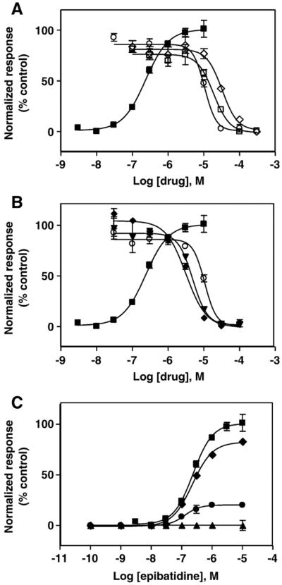
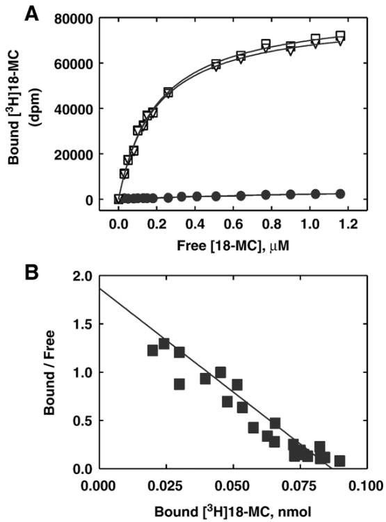
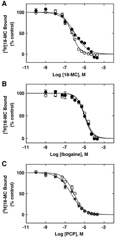
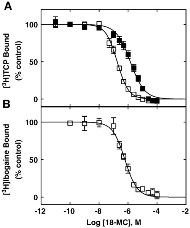
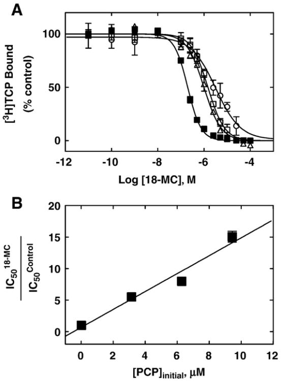
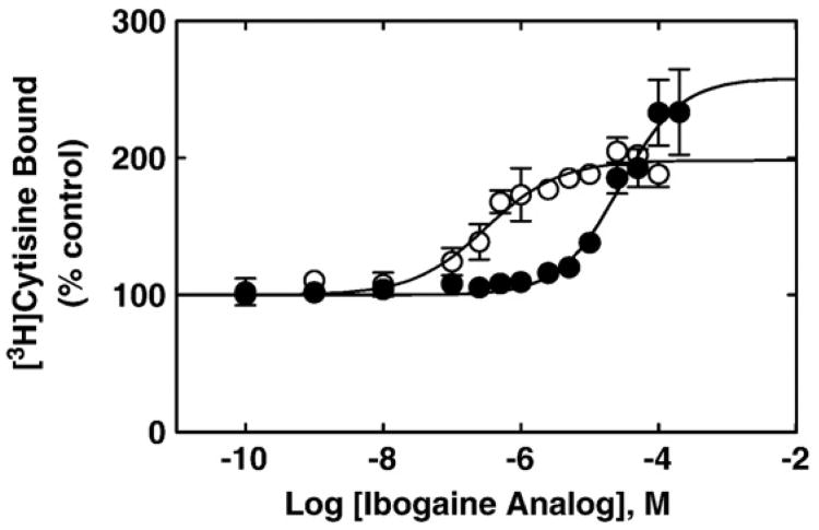
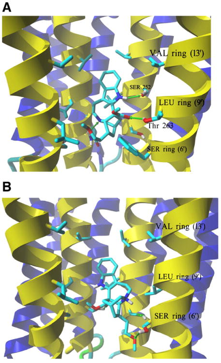
The interaction of 18-methoxycoronaridine (18-MC) with nicotinic acetylcholine receptors (AChRs) was compared with that for ibogaine and phencyclidine (PCP). The results established that 18-MC: (a) is more potent than ibogaine and PCP inhibiting (+/-)-epibatidine-induced AChR Ca(2+) influx. The potency of 18-MC is increased after longer pre-incubation periods, which is in agreement with the enhancement of [(3)H]cytisine binding to resting but activatable Torpedo AChRs, (b) binds to a single site in the Torpedo AChR with high affinity and inhibits [(3)H]TCP binding to desensitized AChRs in a steric fashion, suggesting the existence of overlapping sites. This is supported by our docking results indicating that 18-MC interacts with a domain located between the serine (position 6') and valine (position 13') rings, and (c) inhibits [(3)H]TCP, [(3)H]ibogaine, and [(3)H]18-MC binding to desensitized AChRs with higher affinity compared to resting AChRs. This can be partially attributed to a slower dissociation rate from the desensitized AChR compared to that from the resting AChR. The enthalpic contribution is more important than the entropic contribution when 18-MC binds to the desensitized AChR compared to that for the resting AChR, and vice versa. Ibogaine analogs inhibit the AChR by interacting with a luminal domain that is shared with PCP, and by inducing desensitization.


Figures

Fig. 1
Fig. 1
Effect of 18-methoxycoronaridine (18-MC), ibogaine, and PCP on (±)-epibatidine-induced calcium influx in TE671-hα1β1γδ cells. (A) Increased concentrations of (±)-epibatidine (■) activate the hα1β1γδ AChR with potency EC50 = 0.26 ±0.04μM (nH = 1.23 ±0.06). Subsequently, cells were pre-treated for 5min with several concentrations of 18-MC (○), ibogaine (□), and PCP (◊) followed by addition of 1 μM (±)-epibatidine. (B) Longer pre-treatment increases the inhibitory potency of 18-MC on (±)-epibatidine-induced calcium influx in TE671-hα1β1γδ cells. Pre-treatment periods were: 5 min (○), 4 h (▼), and 24 h (◆), respectively. Responses in both plots were normalized to the maximal (±)-epibatidine response which was set as 100%. The calculated IC50 and nH values are summarized in Table 1. (C) Pre-treatment with 1 (◆), 10 (●), and 100 μM 18-MC (▲), respectively, inhibits (±)-epibatidine-induced calcium influx in TE671-hα1β1γδ cells in a dose dependent and noncompetitive manner. The plots are representative of twenty-seven (■), nine (○), six (□), three (◊) and three (▲,▼,◆,●) determinations, respectively, where the error bars represent the standard deviation (S.D.) values.
Fig. 2
Fig. 2
Equilibrium binding of [3H]18-MC to Torpedo AChRs in the desensitized/CCh-bound state. (A) Total (□), nonspecific (●) (in the presence of 100 μM 18-MC), and specific (○) (total —nonspecific binding) [3H]18-MC binding. Torpedo AChR native membranes (0.5 μM) were suspended in BS buffer, in the presence of 1 mM CCh, and pre-incubated for 30 min at RT. Then, the total volume of the membrane suspensions (total and nonspecific binding) was divided into aliquots and increasing concentrations of [3H]18-MC +18-MC (i.e., 0.07 to 1.2 μM) were added to each tube and incubated for 2 h at RT. Finally, the AChR-bound [3H]18-MC was separated from the free ligand by using the filtration assay described in Section 2.4. (B) Rosenthal-Scatchard plot for [3H] 18-MC specific binding to the Torpedo AChR ion channel. The Kd value (0.23 ±0.04μM) was determined from the negative reciprocal of the slope, according to Eq. (1). The stoichiometric ratio (0.86 ± 0.13 binding sites/AChR) was obtained from the x-intersect (when y = 0) of the plot [B]/[F]versus[B] according to Eq. (1), considering the AChR concentration in the assay. Shown is the combination of two separate experiments.
Fig. 3
Fig. 3
(A) Inhibition of [3H]18-MC binding to Torpedo AChRs in different conforma-tional states by (A) 18-MC, (B) ibogaine, and (C) PCP, respectively. AChR-rich membranes (0.3 μM) were equilibrated (2 h) with 3.6 (●) or 5.5 nM (○) [3H]18-MC, in the presence of 1 mM CCh (●) (desensitized/CCh-bound state) or 1 μM α-BTx (○) (resting/α-BTx-bound state), respectively, and increasing concentrations of the NCA under study. Nonspecific binding was determined in the presence of 50 (●) or 100 μM 18-MC (○), respectively. Each plot is the combination of two separated experiments each one performed in triplicate, where the error bars represent the standard deviation (S.D.) values. From these plots the IC50 and nH values were obtained by nonlinear least-squares fit according to Eq. (2). Subsequently, the Ki values were calculated using Eq. (3). The calculated Ki and nH values are summarized in Table 2.
Fig. 4
Fig. 4
18-MC-induced inhibition of (A) [3H]TCP and (B) [3H]ibogaine binding to Torpedo AChRs in different conformational states. AChR-rich membranes (0.3 μM) were equilibrated (2 h) with 4 nM [3H]TCP in the presence of 1 mM CCh (□) (desensitized/CCh-bound state) or 1 μM α-BTx (■) (resting/α-BTx-bound state), or alternatively with 19 nM [3H]ibogaine in the presence of 1 mM CCh (□), and increasing concentrations of 18-MC. Nonspecific binding was determined in the presence of 100 μM PCP. Each plot is the combination of two separated experiments each one performed in triplicate, where the error bars represent the standard deviation (S.D.) values. From these plots the IC50 and nH values were obtained by nonlinear least-squares fit according to Eq. (2). Subsequently, the Ki values were calculated using Eq. (3). The calculated Ki and nH values are summarized in Table 2.
Fig. 5
Fig. 5
Schild-plot for 18-MC-induced inhibition of [3H]TCP binding to the Torpedo AChR in the desensitized/CCh-bound state. (A) AChR-rich membranes (0.3 μM nAChR) were equilibrated with [3H]TCP, in the presence of 1 mM CCh (desensitized state), at initial PCP concentrations of 0 (control; ■), 3.1 (Δ),6.3 (□),and 9.4μM (○), respectively. The apparent IC50 values were calculated by nonlinear least-squares fit according to Eq. (2). Shown is the average of experiments performed in triplicate. (B) Modified Schild-plot for 18-MC-induced inhibition of [3H]TCP binding. The plot shows a linear relationship with a r2 value of 0.92. Shown is the result of experiments performed in triplicate, where the error bars represent the standard deviation (S.D.) values.
Fig. 6
Fig. 6
Ibogaine analog-induced enhancement of [3H]cytisine binding to Torpedo AChRs in the resting but activatable state. AChR native membranes (0.3 μM nAChR) were equilibrated (30 min) with 7.7 nM [3H]cytisine, and increasing concentrations of ibogaine (●) and 18-MC (○), respectively. Each plot is the combination of 2–3 separated experiments each one performed in triplicate, where the error bars represent the standard deviation (S.D.) values. The apparent EC50 values, obtained according to Eq. (2), were 28±10 (apparent nH=0.99±0.22) and 0.3±0.1 μM (apparent nH= 0.81±0.20) for ibogaine and 18-MC, respectively.
Fig. 7
Fig. 7
Complexes formed between 18-MC and AChR ion channel models obtained by molecular docking. (A) Side view of the lowest energy complex formed between neutral 18-MC and the human muscle AChR ion channel. Receptor subunits are shown in the secondary structure mode (M2 helices, yellow; other transmembrane helices, blue) with residues forming the serine (SER) (position 6′), leucine (LEU) (positions 9′), and valine (VAL) (position 13′) rings, shown explicitly in stick mode. Green arrows indicate hydrogen bonds formed between the pyrrole amino group and the α1-Ser252 residue (position 10′), another between the ligand ionizable amino group and γ-Asn257 at the SER ring, and a third one between the carbonyl of the ester group and β1-Thr263 (position 10′). A similar interaction was obtained for protonated 18-MC. (B) Side view of the lowest energy complex formed between protonated 18-MC and the Torpedo AChR ion channel. The subunit γ was hidden for clarity, and the order of the remaining subunits is (from the left): α1, β1,δ, α1. The ether and ester groups from 18-MC form hydrogen bonds with two Ser residues, each one from the respective α1 and β1 subunits, forming the SER ring. Another strong hydrogen bond interaction is observed between the charged amino group and the same α1-Ser residue. 18-MC interacts with the LEU and VAL rings by additional weaker van der Waals contacts. A similar interaction was obtained for neutral 18-MC.

Similar articles

Cited by

References

    1. Arias HR. Thermodynamics of nicotinic receptor interactions. In: Raffa RB, editor. Drug-Receptor Thermodynamics: Introduction and Applications. John Wiley & Sons, Ltd; USA: 2001. pp. 293–358.
    1. Arias HR. Ligand-gated ion channel receptor superfamilies. In: Arias HR, editor. Biological and Biophysical Aspects of Ligand-Gated Ion Channel Receptor Superfamilies. Research Signpost; Kerala, India: 2006. pp. 1–25.
    1. Arias HR, Bhumireddy P, Bouzat C. Molecular mechanisms and binding site locations for noncompetitive antagonists of nicotinic acetylcholine receptors. Int J Biochem Cell Biol. 2006;38:1254–1276. - PubMed
    1. Albuquerque EX, Pereira EFR, Alkondon A, Rogers SW. Mammalian nicotinic acetylcholine receptors: From structure to function. Physiol Rev. 2009;89:73–120. - PMC - PubMed
    1. Sine SM, Engel AG. Recent advances in Cys-loop receptor structure and function. Nature. 2006;440:448–455. - PubMed

Publication types

LinkOut - more resources