. 2010 Jun 25;636(1-3):198-202.
doi: 10.1016/j.ejphar.2010.03.010. Epub 2010 Mar 19.

Repression of beta-catenin signaling by PPAR gamma ligands

Affiliations

Repression of beta-catenin signaling by PPAR gamma ligands

Desheng Lu et al. Eur J Pharmacol. .

Abstract

Aberrant activation of the Wnt/beta-catenin signaling pathway plays a crucial role in oncogenesis of various human malignancies. It has been demonstrated that there is a direct interaction between beta-catenin and PPAR gamma. Here we examined the effects of fifteen reported PPAR ligands in a reporter gene assay that is dependent on beta-catenin activation of TCF/LEF transcription factors; only the thiazolidinedione PPAR gamma agonists troglitazone, rosiglitazone and pioglitazone, and a non-thiazolidinedione PPAR gamma activator GW1929 inhibited beta-catenin-induced transcription in a PPAR gamma dependent fashion. The results from mammalian one-hybrid experiments showed that functional PPAR gamma was necessary for ligand-dependent inhibition of beta-catenin transactivation. However, a PPAR gamma activator Fmoc-Leu could not repress beta-catenin-mediated signaling and its transactivation activity. These results indicate that activation of PPAR gamma is necessary, but not sufficient, for the beta-catenin antagonistic activity of a PPAR gamma agonist, and that the inhibitory compounds interfere directly with beta-catenin transactivation activity.


Figures

Fig. 1
Fig. 1
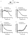
PPARγ agonists repress β-catenin-mediated signaling in a PPARγ dependent manner. (A) A schematic representation of the TOPflash reporter used in this study. TOPflash reporter was transfected into HEK293 cells with expression plasmids for Dvl, RXRα and PPARγ or PPARγEA469 as indicated. After overnight incubation, transfected cells were treated for 24 h with increasing concentrations of troglitazone (B), rosiglitazone (C), GW1929 (D) and Fmoc-Leu (E), after which the reporter gene activity was measured. The results are expressed as % control TOPflash reporter activity ±S.E.M.
Fig. 2
Fig. 2
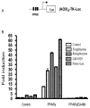
PPARγ agonists stimulate PPRE reporter gene transcription. (A) A schematic representation of the PPRE reporter (AOX)3-TK-Luc. (B) HEK293 cells were transfected with (AOX)3-TK-Luc reporter with or without PPARγ or PPARγEA469, and then were incubated with 5 μM of different PPARγ ligands, as indicated. The fold increase in luciferase activities ±S.E.M. was determined.
Fig. 3
Fig. 3
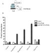
PPARγ agonists induce the interaction of PPARγ with its binding protein PBP. (A) Schematic representation of the mammalian two-hybrid system used in this study. (B) The reporter plasmid UAS-TK-Luc was transfected into HEK293 cells with a VP16-PPARγ expression plasmid, either alone or with VP16-PPARγ and Gal4-PBP. Transfected cells were treated with different PPARγ activators (5 μM each) as indicated. Cells were harvested 24 h after treatment, and then luciferase activities were determined.
Fig. 4
Fig. 4
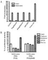
PPARγ agonists repress the transactivation activity of β–catenin in the presence of functional PPARγ. (A) The reporter gene UAS-TK-Luc was transfected into HEK293 cells, together with a Gal4-β-catenin expression plasmid. Then cells were exposed to 5 μM troglitazone, 5 μM rosiglitazone, 5 μM GW1929, 5 μM Fmoc-Leu or vehicle alone for 24 h, harvested, and extracted for determination of luciferase activity. The results are expressed as fold induction of luciferase activity compared to the basal level ±S.E.M. (B) HEK293 cells were transfected with UAS-TK-Luc, the reporter construct, along with expression plasmids for Gal4-β-catenin, RXRα, PPARγ or PPARγEA469, as indicated. After transfection, cells were grown with the PPARγ agonists troglitazone, rosiglitazone, GW1929 and Fmoc-Leu (5 μM each), and assayed for luciferase activity 24 h later. The results are expressed as % control reporter activity ±S.E.M.

Similar articles

Cited by

References

    1. Altiok S, Xu M, Spiegelman BM. PPARgamma induces cell cycle withdrawal: inhibition of E2F/DP DNA-binding activity via down-regulation of PP2A. Genes Dev. 1997;11:1987–1998. - PMC - PubMed
    1. Clevers H. Wnt/beta-catenin signaling in development and disease. Cell. 2006;127:469–480. - PubMed
    1. Crawford HC, Fingleton BM, Rudolph-Owen LA, Goss KJ, Rubinfeld B, Polakis P, Matrisian LM. The metalloproteinase matrilysin is a target of beta-catenin transactivation in intestinal tumors. Oncogene. 1999;18:2883–2891. - PubMed
    1. Debril MB, Renaud JP, Fajas L, Auwerx J. The pleiotropic functions of peroxisome proliferator-activated receptor gamma. Journal of molecular medicine (Berlin, Germany) 2001;79:30–47. - PubMed
    1. Forman BM, Tontonoz P, Chen J, Brun RP, Spiegelman BM, Evans RM. 15-Deoxy-delta 12, 14-prostaglandin J2 is a ligand for the adipocyte determination factor PPAR gamma. Cell. 1995;83:803–812. - PubMed

Publication types

MeSH terms