Review
. 2010 Sep;29(5):428-42.
doi: 10.1016/j.preteyeres.2010.03.002. Epub 2010 Mar 19.

Membrane-binding and enzymatic properties of RPE65

Affiliations
Review

Membrane-binding and enzymatic properties of RPE65

Philip D Kiser et al. Prog Retin Eye Res. 2010 Sep.

Abstract

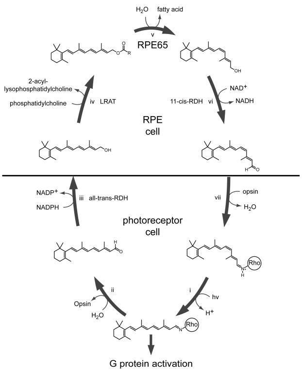
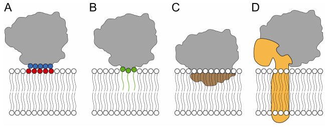
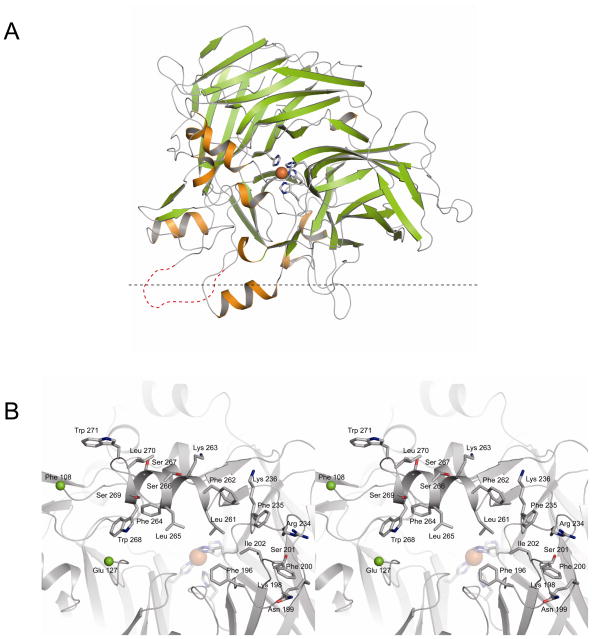
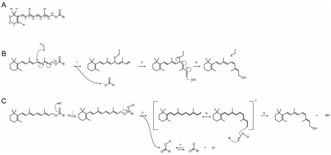
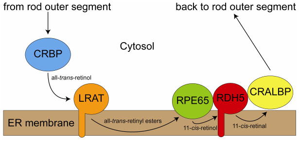
Regeneration of visual pigments is essential for sustained visual function. Although the requirement for non-photochemical regeneration of the visual chromophore, 11-cis-retinal, was recognized early on, it was only recently that the trans to cis retinoid isomerase activity required for this process was assigned to a specific protein, a microsomal membrane enzyme called RPE65. In this review, we outline progress that has been made in the functional characterization of RPE65. We then discuss general concepts related to protein-membrane interactions and the mechanism of the retinoid isomerization reaction and describe some of the important biochemical and structural features of RPE65 with respect to its membrane-binding and enzymatic properties.


Figures

Figure 1
Figure 1
Schematic representation of the retinoid (visual) cycle - Vision begins when light (hv) causes photoisomerization of the 11-cis-retinylidene chromophore of ground-state rhodopsin. Subsequently, the Schiff base linkage loses a proton enabling rhodopsin to activate G proteins (i). After remaining active for a short period of time, the isomerized chromophore is released via hydrolysis, generating free all-trans-retinal and opsin (ii). The all-trans-retinal is enzymatically reduced (iii) and the resultant all-trans-retinol is exported from the rod outer segment to the RPE. Here all-trans-retinol is metabolized by LRAT to produce all-trans-retinyl esters (iv), which can be either stored in retinosomes or further processed. RPE65 is the key enzyme that catalyzes the conversion of all-trans-retinyl esters to 11-cis-retinol (v). 11-cis-Retinol is enzymatically reduced to 11-cis-retinal (vi), which is then transported back to the photoreceptor outer segment where it recombines with opsin to form ground-state rhodopsin (vii). Continuous operation of this cycle is what sustains vision under conditions where rods are primarily active.
Figure 2
Figure 2
Potential mechanisms of RPE65 membrane association - Four potential modes of RPE65-membrane interactions have been proposed based on extraction experiments and other biochemical and biophysical studies. A) Anchoring via electrostatic interactions between RPE65 side chains and the charged headgroups of phospholipids. Blue circles indicate positively charged residues and red circles indicate negatively charged phospholipid moieties. This mode of interaction was proposed based on the results of high-salt and carbonate extraction experiments. B) Anchoring via covalently attached S-palmitoyl groups. Green circles and lines indicate the cysteine and palmitate moieties, respectively. This mode of interaction was proposed on the basis of mass spectrometry experiments. C) Anchoring via direct interactions between hydrophobic side chains (colored brown) and the lipid matrix. The mode of interaction is supported by detergent extraction and phase separation experiments as well as structural and enzymological observations. D) Attachment via interactions with other membrane-bound proteins. The observation that RPE65 appears to form complexes with other RPE microsomal proteins might suggest this mode of interaction. A hypothetical transmembrane protein is shown colored orange. Further details and considerations are discussed in the text
Figure 3
Figure 3
Crystallographic structure of bovine RPE65 - A) The chain adopts a seven bladed beta propeller fold with one face covered by a helical cap. The iron cofactor, shown as an orange sphere, is directly coordinated by four histidine residues that are conserved amongst members of the CCO family. The red dashed line indicates a portion of the chain (residues 109–126) that was unresolved in the final electron density maps. The putative membrane-binding surface is facing down in this panel with the horizontal, black dashed line indicating the hypothetical interface between headgroup and core regions of the membrane. B) Stereoview of the putative membrane-binding surface of RPE65 - The portion of the protein structure facing down in panel A has been rotated 90 degrees around the horizontal axis and is now projects towards the viewer. Segments of the chain that surround the entrance to the active site are enriched in residues that are favored in interfacial (Arg, Lys, Ser, Trp and Tyr) and lipid core regions (Phe, Leu and Ile) of the membrane. Therefore, these regions probably interact extensively with the membrane including the lipid core, consistent with the need for this enzyme to assess membrane-dissolved retinyl esters. The view is approximately down the substrate entry/product exit tunnel with the iron cofactor shown as an orange sphere. The two green spheres indicate the positions of Phe 108 and Glu 127, which flank the unresolved region of the RPE65 chain.
Figure 4
Figure 4
Proposed catalytic mechanisms of retinoid isomerization - Two mechanisms for retinoid isomerization have been proposed that account for the experimentally observed loss of the C15-bound oxygen from the all-trans-retinyl ester during enzymatic processing. A) A retinoid molecule showing the standard numbering scheme. R indicates a saturated hydrocarbon chain B) In this dual SN2-type nucleophilic substitution mechanism a protein associated nucleophile (X) (e.g. a Cys side chain sulfur atom) attacks C11 (i) resulting in the loss of the ester moiety via alkyl-oxygen cleavage and the formation of an intermediate (ii) with a freely rotatable C11-C12 single bond. In the second step (iii) another nucleophile, presumably a hydroxide ion, attacks C15 when the intermediate is in a cis-like conformation, resulting in loss of the C11-bound protein group and restoration of the conjugated double bond system. C) In this SN1-type nucleophilic substitution mechanism, the rate-limiting step, i.e. loss of the ester via alkyl-oxygen cleavage, is catalyzed by a Lewis acid (X+) such as a metal ion or a proton (i). Dissociation of the ester (ii) and the generation of a resonance-stabilized carbocation (iii) lowers the polyene chain carbon-carbon bond order and the activation energy needed for trans to cis isomerization. After rotation to a cis-like conformation, a water molecule, which is either concomitantly or subsequently deprotonated by a base (B), attacks C15, restoring the conjugated double bond system in the 11-cis configuration (iv). Dissociation of the ester leaving group regenerates the Lewis acid catalyst (v). In both mechanisms the thermodynamic energy needed for the endergonic isomerization reaction may derive from the ester hydrolysis or from a downstream process such as interaction of the 11-cis products with retinoid-binding proteins.
Figure 5
Figure 5
Proposed flow of retinoids within the RPE - In this scheme, all-trans-retinol from the rod outer segments, bound by cellular retinol-binding protein in the RPE cytosol, is presented to LRAT for esterification. The resulting esters diffuse through the membrane to RPE65, which catalytically isomerizes them to 11-cis-retinol. Based on the presence of only one suitable tunnel for retinoid passage observed in the RPE65 structure, we propose that the 11-cis-retinol product reenters the membrane and diffuses to RDH5. The 11-cis-retinal product is taken up either from the membrane or directly from RDH5 by CRALBP and transported to the RPE plasma membrane where it is released from the RPE cell and is transported back to the rod outer segment by an incompletely understood process.
Figure 6
Figure 6
Stereoview of the RPE65 interior cavity - The internal cavity of RPE65 housing the iron cofactor is lined with several aromatic residues possibly involved in stabilizing a carbocation intermediate of the retinoid isomerization reaction. The blue mesh delineates the boundaries of this cavity. The carbon atoms of Trp, Tyr and Phe residues are colored orange and the iron cofactor is displayed as an orange sphere.
Figure 7
Figure 7
Stereoview of the RPE65 iron center and surrounding residues - The iron inner coordination sphere consists of four histidine residues arranged in a distorted octahedral geometry. One or two of the remaining sites in the octahedral geometry are filled by non-protein electron density of uncertain origin (not shown but discussed in the text). The Val 134 side chain may block access to one of the iron coordination sites. His residues 241, 313 and 527 hydrogen bond with Glu residues 148, 417 and 469, respectively. Additionally, there are hydrogen bonding interactions between these Glu residues and Tyr 239, Trp 331 and Arg 44, respectively. With the exception of Val 134, all of these residues are known to be critical for RPE65 activity based on data from either from in vitro isomerase activity assays or LCA patients with RPE65 mutations. This arrangement suggests a highly tuned metal-binding site that is extremely susceptible to perturbation by amino acid substitutions. The iron cofactor and a water molecule are shown as brown and red spheres, respectively. Dashed lines indicate interactions, either hydrogen bonds or coordinate bonds, between atoms.

Similar articles

Cited by

References

    1. Abe I, Prestwich GD. Molecular cloning, characterization, and functional expression of rat oxidosqualene cyclase cDNA. Proc Natl Acad Sci U S A. 1995;92:9274–9278. - PMC - PubMed
    1. Alberts B, Johnson A, Lewis J, Raff M, Roberts K, Walter P. Molecular Biology of the Cell. 4. Garland Science; New York: 2002.
    1. Balliano G, Viola F, Ceruti M, Cattel L. Characterization and partial purification of squalene-2,3-oxide cyclase from Saccharomyces cerevisiae. Arch Biochem Biophys. 1992;293:122–129. - PubMed
    1. Barry RJ, Canada FJ, Rando RR. Solubilization and partial purification of retinyl ester synthetase and retinoid isomerase from bovine ocular pigment epithelium. J Biol Chem. 1989;264:9231–9238. - PubMed
    1. Batten ML, Imanishi Y, Maeda T, Tu DC, Moise AR, Bronson D, Possin D, Van Gelder RN, Baehr W, Palczewski K. Lecithin-retinol acyltransferase is essential for accumulation of all-trans-retinyl esters in the eye and in the liver. J Biol Chem. 2004;279:10422–10432. - PMC - PubMed

Publication types

LinkOut - more resources