. 2010 Jun 5;401(2):236-47.
doi: 10.1016/j.virol.2010.02.019. Epub 2010 Mar 21.

Lack of complex N-glycans on HIV-1 envelope glycoproteins preserves protein conformation and entry function

Affiliations

Lack of complex N-glycans on HIV-1 envelope glycoproteins preserves protein conformation and entry function

Dirk Eggink et al. Virology. .

Erratum in

  • Virology. 2010 Oct 10;406(1):162-3

Abstract

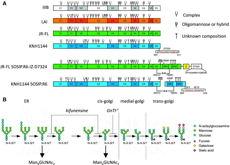
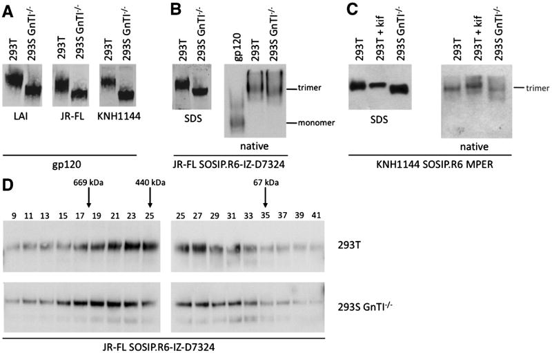
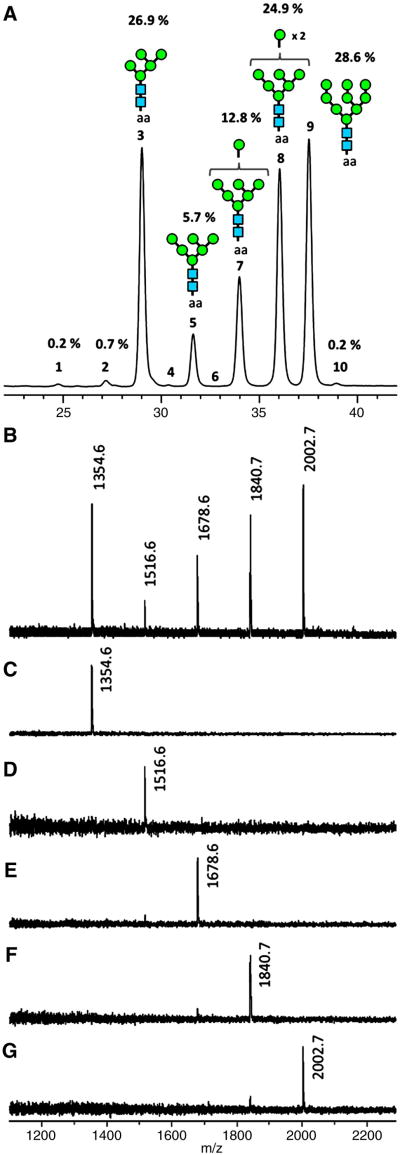
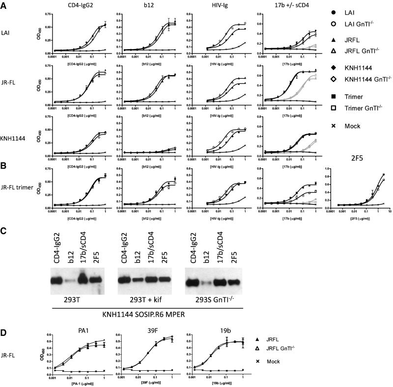
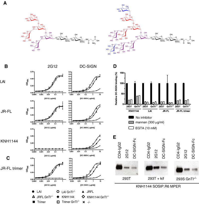
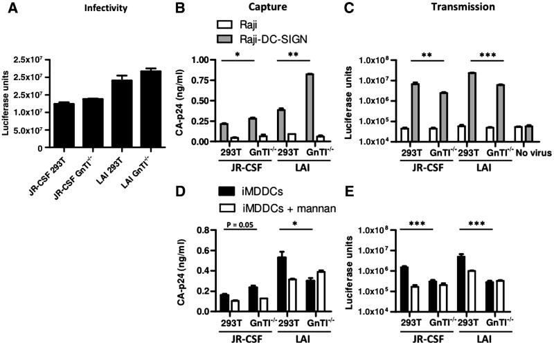
The HIV-1 envelope glycoprotein complex (Env) is the focus of vaccine development aimed at eliciting humoral immunity. Env's extensive and heterogeneous N-linked glycosylation affects folding, binding to lectin receptors, antigenicity and immunogenicity. We characterized recombinant Env proteins and virus particles produced in mammalian cells that lack N-acetylglucosaminyltransferase I (GnTI), an enzyme necessary for the conversion of oligomannose N-glycans to complex N-glycans. Carbohydrate analyses revealed that trimeric Env produced in GnTI(-/-) cells contained exclusively oligomannose N-glycans, with incompletely trimmed oligomannose glycans predominating. The folding and conformation of Env proteins was little affected by the manipulation of the glycosylation. Viruses produced in GnTI(-/-) cells were infectious, indicating that the conversion to complex glycans is not necessary for Env entry function, although virus binding to the C-type lectin DC-SIGN was enhanced. Manipulating Env's N-glycosylation may be useful for structural and functional studies and for vaccine design.


Figures

Figure 1
Figure 1
Study design. (A) Schematic of the gp120 and gp140 proteins used in this study. Constructs based on the sequences from LAI, JR-FL and KNH1144 are indicated in orange, green and blue, respectively. KNH1144 gp120 was modified to reconstitute the epitope for D7324 in the C5 domain (substitutions: R500K, R502K and G507Q). JR-FL SOSIP.R6-IZ-D7324 gp140 and KNH1144 SOSIP.R6 MPER gp140 contain several modifications that have mostly been described elsewhere, including the A501C and T605C substitutions to create the SOS disulfide bond (Binley et al., 2000); the I559P substitution to promote trimerization (Sanders et al., 2002b); the GCN4-based isoleucine zipper (IZ) introduction to promote trimerization (Harbury, Kim, and Alber, 1994; Yang et al., 2000a; Yang et al., 2000b) followed by an D7324 epitope tag; the hexa-arginine (R6) cleavage site to enhance cleavage (Binley et al., 2002); and the MPER substitutions, A662E, G664D, N668S and T671N (indicated in red) to introduce the 2F5 and 4E10 epitopes into KNH1144 SOSIP.R6 gp140 (Dey et al., 2009). The N-linked glycan sites on gp120 and gp140 proteins produced in wild type mammalian cells are designated as oligomannose or complex, based on experimental determinations using IIIB gp120 (Leonard et al., 1990). It is assumed that the glycans present at analogous sites are processed similarly on the other gp120s, but we note that the N-glycan type (oligomannose vs complex) present at some sites depends on the study and isolate used (Cutalo, Deterding, and Tomer, 2004; Leonard et al., 1990; Zhu et al., 2000). Furthermore, some sites can be unoccupied in a only subset of molecules, or be occupied by both complex and oligomannose sugars (Cutalo, Deterding, and Tomer, 2004; Zhu et al., 2000). Sites that are present on LAI, JR-FL or KNH1144 gp120, but not on IIIB gp120, are designated as being of unknown glycan composition. The glycans on gp41 have not been characterized. (B) Schematic of the mammalian N-linked glycosylation pathway with the blocks in 293S GnTI-/- cells and in 293T cells treated with kifunensine indicated. The cellular sites at which processing occurs are also indicated.
Figure 2
Figure 2
Expression of gp120 and gp140 in 293T cells and in 293S cells lacking the GnTI enzyme. (A) SDS-PAGE analysis of LAI, JR-FL and KNH1144 gp120s expressed transiently in 293T or 293S GnTI-/- cells. (B) SDS-PAGE and BN-PAGE analysis of uncleaved JR-FL SOSIP.R6-IZ-D7324 gp140, expressed in both cell types. (C) SDS-PAGE and BN-PAGE analysis of cleaved KNH1144 SOSIP.R6 MPER gp140 expressed in the presence of furin, in either 293T cells with or without kifunensine, or in 293S GnTI-/- cells. (D) Size exclusion chromatography analysis of uncleaved JR-FL SOSIP.R6-IZ gp140 trimers produced in 293T or 293S GnTI-/- cells and fractionated using a Superose-6 column. The fractions (indicated on top) were analyzed by SDS-PAGE and western blot. The peak fractions of the standard protein thyroglobulin (669 kD), ferritin (440 kD) and BSA (67 kD) are indicated.
Figure 3
Figure 3
Glycosylation analysis of gp140 trimers produced in the absence of GnTI. (A) HILIC chromatogram and (B) MALDI-TOF MS spectrum of enzymatically released, 2-AA-labeled N-glycans of KNH1144 trimers from 293S GnTI-/- cells. (C-G) MALDI-TOF MS spectra of RP-LC fractions 3,5,7,8 and 9, respectively.
Figure 4
Figure 4
Antigenic properties of gp120 and gp140 proteins that lack complex N-glycans. ELISA analysis of the binding of HIV-Ig, b12, 17b (in the absence of sCD4: grey lines and symbols; in the presence of sCD4: black lines and symbols) and CD4-IgG2 to: (A) LAI, JR-FL or KNH1144 gp120 proteins expressed in 293T cells or in 293S GnTI-/- cells; (B) uncleaved JR-FL SOSIP.R6-IZ-D7324 gp140 expressed in the same two cell types. (C) Immunoprecipitation analysis of cleaved KNH1144 SOSIP.R6 MPER gp140 expressed in 293T cells with furin and with or without kifunensine, or in 293S GnTI-/- cells. The Env proteins were precipitated with CD4-IgG2, b12 or 2F5, then analyzed by SDS-PAGE and western blotting. (D) ELISA analysis of the binding of the V3 MAbs 39F, 19b, and PA1 with JR-FL gp120 expressed in 293T or GnTI-/- cells.
Figure 5
Figure 5
Interactions with 2G12 and DC-SIGN of gp120 and gp140 proteins lacking complex N-glycans. (A) Chemical structures of Man5GlcNAc2 (left) and Man9GlcNAc2 (right) N-glycans commonly found on proteins expressed in 293S GnTI-/- cells, and in kifunensine-treated 293T cells, respectively (Elbein et al., 1990; Reeves et al., 2002). The individual mannoses that are involved in 2G12 binding (blue) and DC-SIGN binding (core binding site: red; additional contacts: burgundy and blue) are indicated (Calarese et al., 2005; Calarese et al., 2003; Feinberg et al., 2007; Feinberg et al., 2001; Guo et al., 2004; Scanlan et al., 2002; van Liempt et al., 2006). The sugar linkages are also shown. (B) Binding of 2G12 and DC-SIGN to three different gp120s produced in 293T or 293S GnTI-/- cells. (C) Binding of 2G12 and DC-SIGN to uncleaved JR-FL SOSIP.R6-IZ-D7324 gp140 trimers. (D) Inhibition of DC-SIGN binding to LAI, JR-FL and KNH1144 gp120 and JR-FL SOSIP.R6-IZ-D7324 gp140 by mannan or EGTA. (E) Binding of cleaved KNH1144 SOSIP.R6 MPER gp140 to 2G12 and DC-SIGN, analyzed by immunoprecipitation.
Figure 6
Figure 6
Virus produced in 293S GnTI-/- cells is infectious and can be captured efficiently by DC-SIGN expressing cells. (A) Infectivity of LAI and JR-CSF virus produced in 293T and 293S GnTI-/- cells. Equal amounts of the various virus stocks (1 ng of CA-p24 antigen) were used to infect TZM-bl reporter cells. (B) Capture of LAI and JR-CSF viruses produced in 293T and 293S GnTI-/- cells by Raji-DC-SIGN cells. (C) Transmission of GnTI-/- produced virus from Raji-DC-SIGN cells. (D) Capture of LAI and JR-CSF viruses produced in 293T and 293S GnTI-/- cells by DC. (E) Transmission of GnTI-/- produced virus from DC to TZM-bl reporter cells. * represents P < 0,05; ** P < 0,005 and *** P < 0,0005.

Similar articles

Cited by

References

    1. Allaway GP, Davis-Bruno KL, Beaudry GA, Garcia EB, Wong EL, Ryder AM, Hasel KW, Gauduin MC, Koup RA, McDougal JS, et al. Expression and characterization of CD4-IgG2, a novel heterotetramer that neutralizes primary HIV type 1 isolates. AIDS Res Hum Retroviruses. 1995;11(5):533–9. - PubMed
    1. Banerjee K, Andjelic S, Klasse PJ, Kang Y, Sanders RW, Michael E, Durso RJ, Ketas TJ, Olson WC, Moore JP. Enzymatic removal of mannose moieties can increase the immune response to HIV-1 gp120 in vivo. Virology. 2009;389(1-2):108–21. - PMC - PubMed
    1. Beddows S, Kirschner M, Campbell-Gardener L, Franti M, Dey AK, Iyer SP, Maddon PJ, Paluch M, Master A, Overbaugh J, VanCott T, Olson WC, Moore JP. Construction and characterization of soluble, cleaved, and stabilized trimeric Env proteins based on HIV type 1 Env subtype A. AIDS Res Hum Retroviruses. 2006;22(6):569–79. - PubMed
    1. Binley JM, Ban YA, Crooks ET, Eggink D, Osawa K, Schief WR, Sanders RW. Role of Complex Carbohydrates in Human Immunodeficiency Virus Type 1 Infection and Resistance to Antibody Neutralization. 2010 Submitted for publication. - PMC - PubMed
    1. Binley JM, Ngo-Abdalla S, Moore P, Bobardt M, Chatterji U, Gallay P, Burton DR, Wilson IA, Elder JH, de Parseval A. Inhibition of HIV Env binding to cellular receptors by monoclonal antibody 2G12 as probed by Fc-tagged gp120. Retrovirology. 2006;3:39. - PMC - PubMed

Publication types

MeSH terms

Substances