. 2010 Jun;31(17):4657-71.
doi: 10.1016/j.biomaterials.2010.01.138. Epub 2010 Mar 21.

Synergistic effects of tethered growth factors and adhesion ligands on DNA synthesis and function of primary hepatocytes cultured on soft synthetic hydrogels

Affiliations

Synergistic effects of tethered growth factors and adhesion ligands on DNA synthesis and function of primary hepatocytes cultured on soft synthetic hydrogels

Geeta Mehta et al. Biomaterials. 2010 Jun.

Abstract

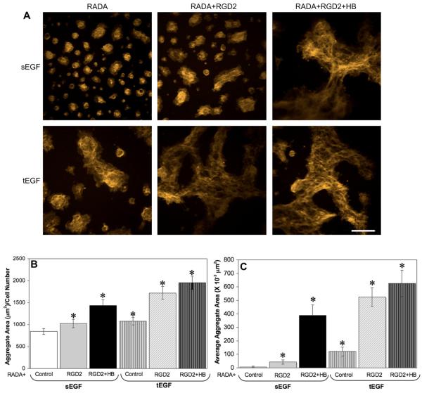
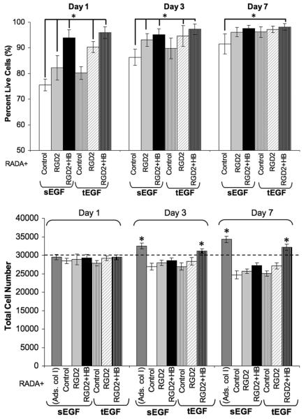
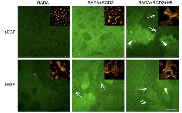
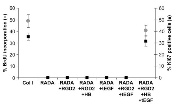
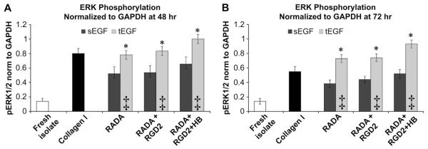
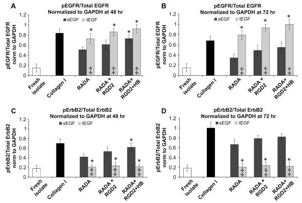
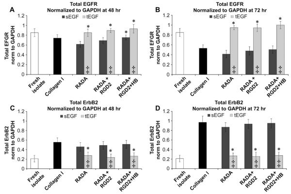
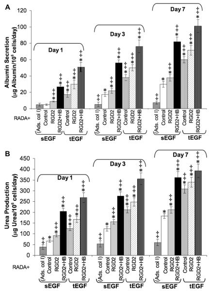
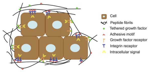
The composition, presentation, and spatial orientation of extracellular matrix molecules and growth factors are key regulators of cell behavior. Here, we used self-assembling peptide nanofiber gels as a modular scaffold to investigate how fibronectin-derived adhesion ligands and different modes of epidermal growth factor (EGF) presentation synergistically regulate multiple facets of primary rat hepatocyte behavior in the context of a soft gel. In the presence of soluble EGF, inclusion of dimeric RGD and the heparin binding domain from fibronectin (HB) increased hepatocyte aggregation, spreading, and metabolic function compared to unmodified gels or gels modified with a single motif, but unlike rigid substrates, gels failed to induce DNA synthesis. Tethered EGF dramatically stimulated cell aggregation and spreading under all adhesive ligand conditions and also preserved metabolic function. Surprisingly, tethered EGF elicited DNA synthesis on gels with RGD and HB. Phenotypic differences between soluble and tethered EGF stimulation of cells on peptide gels are correlated with differences in expression and phosphorylation the EGF receptor and its heterodimerization partner ErbB2, and activation of the downstream signaling node ERK1/2. These modular matrices reveal new facets of hepatocellular biology in culture and may be more broadly useful in culture of other soft tissues.


Figures

Fig. 1
Fig. 1
Self-assembling peptide gels functionalized with the cell-binding domain and heparin binding domain of fibronectin and tethered EGF influence hepatocyte morphology and adhesion. A) Representative fluorescent actin cytoskeleton images (5× objective) for hepatocytes on RADA, RADA + RGD2, RADA + RGD2 + HB peptide gels with soluble EGF or tEGF, 3 days after initial seeding (Scale bar = 400 μm). B) Quantification of average cell aggregate area at day 3 on peptide gels, and C) Quantification of average cell area at day 3 on peptide gels expressed as ratio of total aggregate area and total nuclei counted. Fluorescent actin and DAPI images of each peptide gel sample (seeded at 100,000 cells/cm2) were taken in 4–5 fields at each time point. * indicates statistically significant difference from RADA with soluble EGF, p < 0.05, n > 3.
Fig. 2
Fig. 2
Adhesion ligands and tethered growth factor modulate hepatocyte viability on self-assembling peptides for seven days in culture. A) Quantification of cell viability expressed as percentage live cells as a function of gel composition, B) Total cell numbers as a function of time in culture. Hepatocytes on RADA, RADA + RGD2 RADA + RGD2 + HB peptide gels with soluble EGF or tEGF (seeded at 65,000 cells/cm2) were stained with EtBr and DAPI at each time point. Images representing four random fields were taken on each peptide gel sample. Cell viability was defined as the fraction of non-EtBr stained cells in the field. Except adsorbed collagen I (ads. col I), all peptide gels contained RADA. * indicates statistically significant difference from RADA with soluble EGF at a specific day, p < 0.05, n > 3.
Fig. 3
Fig. 3
Inclusion of adhesion ligands and tethered growth factor instruct hepatocyte to secrete and accumulate matrix after 3 days in culture on self-assembling peptide gels. Representative images of fibronectin and actin (inset) on RADA, RADA + RGD2, RADA + RGD2 + HB peptide gels with soluble EGF or tEGF 3 days after initial seeding at 100,000 cells/cm2 (Scale bar = 400 mm, 5× objective). Cells were fixed, permeabilized and stained with anti-rat fibronectin and four random fields were imaged on each peptide gel sample. Arrows indicate intracellular staining for fibronectin and arrowheads point to extracellular fibrils of fibronectin.
Fig. 4
Fig. 4
Primary hepatocytes require presence of RGD, HB and tEGF on peptide gels in order to synthesize DNA. Quantification of DNA synthesis by BrdU incorporation and active cell cycle marker Ki67. Hepatocytes (seeded at 65,000 cells/cm2) were cultured on RADA, RADA + RGD2 and RADA + RGD2 + HB peptide gels with soluble EGF or tEGF and adsorbed collagen I (Col I). DNA synthesis was quantified by immunofluorescence staining as the percentage of total nuclei incorporating BrdU (48–72 h) or as the percentage of total nuclei staining positive for Ki67 (at 48 h). We observed no DNA synthesis at any time point on other peptide gel (RADA, RADA + RGD2, RADA + RGD2 + HB all with soluble EGF and RADA + tEGF, RADA + RGD2 + tEGF).
Fig. 5
Fig. 5
Addition of tEGF to peptide gels with varying adhesion ligands leads to sustained ERK phosphorylation in hepatocytes. ERK phosphorylation (normalized to GAPDH) at A) 48 h, B) 72 h. Hepatocytes were cultured on RADA, RADA + RGD2 and RADA + RGD2 + HB peptide gels with soluble EGF or tEGF or tEGF (seeded at 100,000 cells/cm2). Lysates were made after defined time-points and pERK levels were quantified by immunoprecipitation. * indicates statistically significant difference from RADA with soluble EGF, ‡ indicates statistically significant difference between soluble and tethered for a particular peptide gel composition, p < 0.05, n > 2.
Fig. 6
Fig. 6
Tethered EGF stimulates sustained EGFR phosphorylation and downregulates ErBB2 phosphorylation in hepatocytes on peptide gels. EGFR phosphorylation (normalized to total EGFR and GAPDH) at A) 48 h, B) 72 h, and ErbB2 phosphorylation (normalized to total ErbB2 and GAPDH) at C) 48 h, D) 72 h. Hepatocytes were cultured on peptide gels with soluble EGF or tEGF or tEGF (seeded at 100,000 cells/cm2). Lysates were made after defined time-points and pEGFR levels were quantified by immunoprecipitation. * indicates statistically significant difference from RADA with soluble EGF, ‡ indicates statistically significant difference between soluble and tethered for a particular peptide gel composition, p < 0.05, n > 2.
Fig. 7
Fig. 7
Tethered EGF maintains total EGFR and downregulates ErbB2 receptor levels on peptide gels. Total EGFR (normalized to GAPDH) at A) 48 h, B) 72 h, and total ErbB2 (normalized to GAPDH) at C) 48 h, D) 72 h. Hepatocytes were cultured on peptide gels with soluble EGF or tEGF or tEGF (seeded at 100,000 cells/cm2). Lysates were made after defined time-points and pEGFR levels were quantified by immunoprecipitation. * indicates statistically significant difference from RADA with soluble EGF, ‡ indicates statistically significant difference between soluble and tethered for a particular peptide gel composition, p < 0.05, n > 2.
Fig. 8
Fig. 8
Maintenance of metabolic function improves with functionalization of peptide gels with adhesion ligands and growth factor tethering. A) Albumin secretion, B) Urea production. Conditioned media from hepatocyte cultures on peptide gels and adsorbed collagen I (seeded at 100,000 cells/cm2) were collected, and urea and albumin were quantified using standard assay kits. Culture medium was replaced 48 h prior to collection on the indicated days. Samples, standards and controls were tested in duplicates. ‡ indicates statistically significant difference from RADA, p < 0.05, n > 3, Except adsorbed collagen I (ads. col I), all peptide gels contained RADA. * indicates statistically significant difference from adsorbed collagen I, p < 0.05, n > 3.
Fig. 9
Fig. 9
Self-assembling peptide gels modified with adhesive ligands and tethered growth factors may provide adequate microenvironment to various cell types. The peptide backbone can be modified with a variety of adhesive modifications suitable to the integrin repertoire of any cell type. Further, inclusion of a biotin modification on the peptide backbone allows any biotinylated growth factor to be tethered to the gel. Given the stiffness of the peptide gels are physiological for many tissues, these gels can be modified to suit a wide array of cell types forming a chemically and mechanically controlled microenvironment.

Similar articles

Cited by

References

    1. Griffith LG, Swartz MA. Capturing complex 3D tissue physiology in vitro. Nat Rev Mol Cell Biol. 2006;7(3):211–24. - PubMed
    1. Davis ME, Hsieh PC, Takahashi T, Song Q, Zhang S, Kamm RD, et al. Local myocardial insulin-like growth factor 1 (IGF-1) delivery with biotinylated peptide nanofibers improves cell therapy for myocardial infarction. Proc Natl Acad Sci U S A. 2006;103(21):8155–60. - PMC - PubMed
    1. Ellis-Behnke RG, Liang YX, You SW, Tay DK, Zhang S, So KF, et al. Nano neuro knitting: peptide nanofiber scaffold for brain repair and axon regeneration with functional return of vision. Proc Natl Acad Sci U S A. 2006;103(13):5054–9. - PMC - PubMed
    1. Horii A, Wang X, Gelain F, Zhang S. Biological designer self-assembling peptide nanofiber scaffolds significantly enhance osteoblast proliferation, differentiation and 3-D migration. PloS One. 2007;2(2):e190, 1–9. - PMC - PubMed
    1. Kisiday J, Jin M, Kurz B, Hung H, Semino C, Zhang S, et al. Self-assembling peptide hydrogel fosters chondrocyte extracellular matrix production and cell division: implications for cartilage tissue repair. Proc Natl Acad Sci U S A. 2002;99(15):9996–10001. - PMC - PubMed

Publication types

MeSH terms