Review
. 2010 May;46(7):1181-8.
doi: 10.1016/j.ejca.2010.02.027. Epub 2010 Mar 19.

Extracellular matrix: a gatekeeper in the transition from dormancy to metastatic growth

Affiliations
Review

Extracellular matrix: a gatekeeper in the transition from dormancy to metastatic growth

Dalit Barkan et al. Eur J Cancer. 2010 May.

Abstract

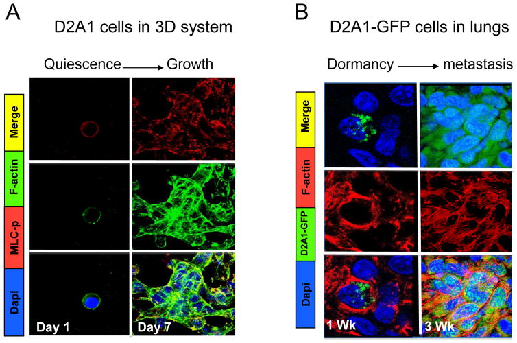
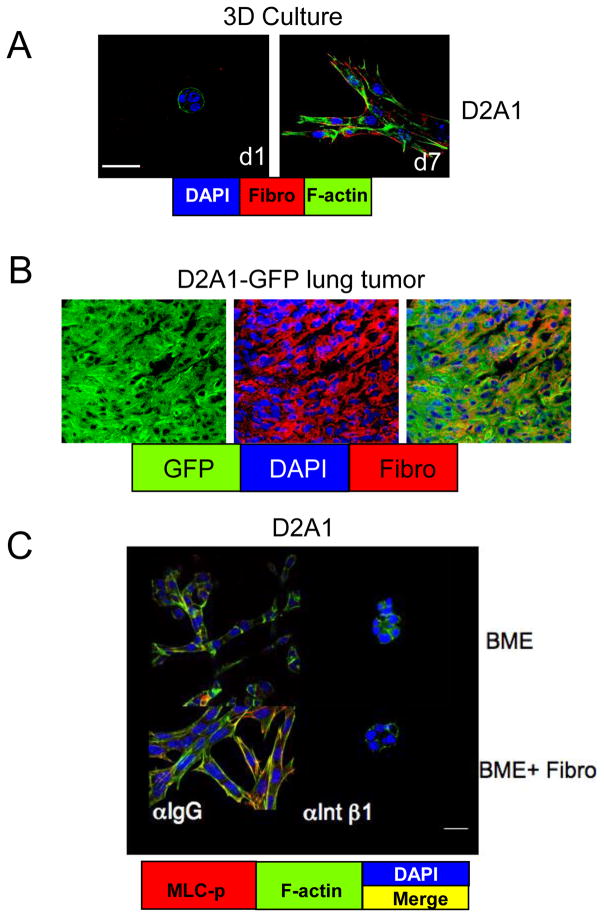
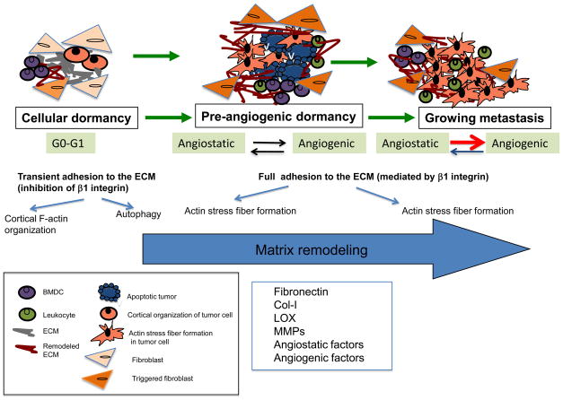
Metastases can develop after apparently successful treatment of a primary tumour, sometimes following a period of tumour dormancy that can last for years. However, factors that regulate metastatic tumour dormancy remain poorly understood. Here we review the potential contribution of interactions between tumour cells and the microenvironment in metastatic sites, in regulating tumour dormancy vs. metastatic growth. We focus particularly on the potential role of the extracellular matrix (ECM) in regulating maintenance and release from dormancy. Tumour cells that fail to properly adhere to the ECM may enter a state of dormancy. The molecular and physical composition of the ECM can be affected by tumour cells themselves, as well as multiple stromal cell types. The roles of integrins, fibronectin, and collagen are discussed, as are factors that can change the ECM. A better understanding of the molecular details of the crosstalk between tumour cells and the ECM in secondary sites, and how these regulate the dormant state, may lead to improved therapeutic strategies to induce or maintain disseminated tumour cells in a dormant state, or alternatively to successfully eradicate dormant cells.


Conflict of interest statement

Conflict of Interest Statement

None declared.

Figures

Figure 1
Figure 1. Cytoskeletal reorganization and formation of actin stress fibers during the switch from dormancy to metastatic growth
A) Confocal microscopy image (Magnification x63) of D2A1 cells cultured in the 3D basement membrane extract (BME) system stained with DAPI (blue), antibody against the phosphorylated form of myosin light chain (MLC-p) (red), and for F-actin (green) at day 1 (d1) and day 7 (d7). Quiescent D2A1 cells displayed cortical F-actin and MLC-p staining on day 1, whereas, during their transition from quiescence to growth (days 4–7) D2A1 cells formed actin stress fibers with elevated expression of MLC-p. B) Frozen sections of the lungs removed from mice injected with D2A1 cells stably expressing GFP (green) at the indicated time points and stained for F-actin (red) and DAPI (blue). One week (1 Wk) post-injection, D2A1-GFP cells reside as solitary dormant cells with cortical staining for F-actin while by week 3 (3 Wk) the metastatic lesion from the D2A1-GFP cell line formed actin stress fibers. Modified from Barkan et al.(32)
Figure 2
Figure 2. Fibronectin expressed by metastatic D2A1 cells signals through β1 integrin leading to transition from dormancy to growth
A–C) Confocal images (magnification x63). A) D2A1 cells cultured on 3D BME express fibronectin during their transition from quiescence to growth (red staining) (day 7). B) Frozen section of lung lesions of D2A1-GFP cells (green) stained for fibronectin (red). C) Treatment of D2A1 cells with a neutralizing antibody against β1 integrin, for 6 days, led to inhibition of MLC-p (staining red), loss of actin stress fiber formation (staining green) and its co-localization with MLC-p (staining yellow) and proliferation of D2A1 cells cultured either on 3D BME or on 3D BME supplemented with fibronectin (BME+Fibro). Treatment with control IgG antibody had no effect. White bar = 20 μm. Modified from Barkan et al.(32)
Figure 3
Figure 3. ECM remodeling by stromal cells and escape from tumor dormancy
The crosstalk between the ECM at the secondary site with the disseminated tumor cell may decide the tumor cell’s fate. Solitary tumor cells that fail to properly adhere to the ECM may enter a state of dormancy activating long-term survival programs such as regulated autophagy. In order to subsequently escape tumor dormancy tumor cells need to fully engage with the ECM components via integrin receptor(s), inducing downstream signaling leading to cytoskeletal reorganization and proliferation. Fibroblasts as well as the tumor cells may remodel the ECM to induce a permissive microenvironment for the tumor cell growth. Changes in the ECM components such as production and organization of Col-I and fibronectin may lead also to the establishment of the pre-metastatic niche by BMDC, leading to the transition from dormancy to growth. After exiting quiescence, the tumor cell mass can enter pre-angiogenic dormancy due to release of angiostatic fragments from the ECM by the MMPs of leukocytes. The angiogenic switch will occur once angiogenic factors are released from the ECM by stromal MMPs, resulting in metastatic growth.

Similar articles

Cited by

References

    1. Aguirre-Ghiso JA. Models, mechanisms and clinical evidence for cancer dormancy. Nat Rev Cancer. 2007;7(11):834–46. - PMC - PubMed
    1. Pantel K, Brakenhoff RH. Dissecting the metastatic cascade. Nat Rev Cancer. 2004;4(6):448–56. - PubMed
    1. Naumov GN, MacDonald IC, Chambers AF, et al. Solitary cancer cells as a possible source of tumour dormancy? Semin Cancer Biol. 2001;11(4):271–6. - PubMed
    1. Townson JL, Chambers AF. Dormancy of solitary metastatic cells. Cell Cycle. 2006;(16):1744–50. - PubMed
    1. Chambers AF, Groom AC, MacDonald IC. Dissemination and growth of cancer cells in metastatic sites. Nat Rev Cancer. 2002;2(8):563–72. - PubMed

Publication types