

Immune surveillance of the maternal/fetal interface: controversies and implications
- PMID: 20304670
- PMCID: PMC2892024
- DOI: 10.1016/j.tem.2010.02.003
Immune surveillance of the maternal/fetal interface: controversies and implications
Abstract
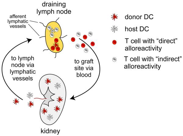
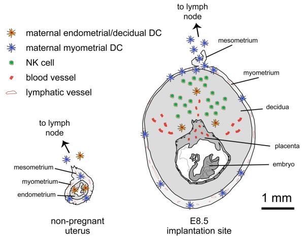
How the fetal 'allograft' avoids rejection during pregnancy remains a major unresolved immunological paradox. Recent work has suggested that fetomaternal tolerance is in fact maintained by a number of redundant mechanisms, but their relative importance has remained poorly defined. In this paper, I discuss an emerging controversy regarding the ability of maternal T cells to mediate fetal rejection at a time when they appear to be ignorant of fetal and placental antigens. This paradox within a paradox highlights two major research directions in the field of reproductive immunology that, when ultimately reconciled, promise to give significant insight into mechanisms of impaired fertility and compromised fetal and maternal health.
Copyright 2010 Elsevier Ltd. All rights reserved.
Figures
Similar articles
-
Tolerance to the foetal allograft?Am J Reprod Immunol. 2010 Jun;63(6):624-36. doi: 10.1111/j.1600-0897.2010.00832.x. Epub 2010 Mar 29. Am J Reprod Immunol. 2010. PMID: 20367624 Review.
-
Pregnancy immune tolerance at the maternal-fetal interface.Int Rev Immunol. 2020;39(6):247-263. doi: 10.1080/08830185.2020.1777292. Epub 2020 Jun 12. Int Rev Immunol. 2020. PMID: 32530719
-
T cell behavior at the maternal-fetal interface.Int J Dev Biol. 2014;58(2-4):189-98. doi: 10.1387/ijdb.140054ae. Int J Dev Biol. 2014. PMID: 25023685 Free PMC article. Review.
-
Immunology of the maternal-placental interface in normal pregnancy.Semin Perinatol. 1991 Jun;15(3):196-205. Semin Perinatol. 1991. PMID: 1718044 Review. No abstract available.
-
Maternal and fetal immune responses during pregnancy.Exp Clin Immunogenet. 1993;10(2):85-102. Exp Clin Immunogenet. 1993. PMID: 8251183 Review.
Cited by
-
Dendritic cell function at the maternal-fetal interface.Expert Rev Clin Immunol. 2011 Sep;7(5):593-602. doi: 10.1586/eci.11.52. Expert Rev Clin Immunol. 2011. PMID: 21895472 Free PMC article.
-
Clinical chorioamnionitis at term V: umbilical cord plasma cytokine profile in the context of a systemic maternal inflammatory response.J Perinat Med. 2016 Jan;44(1):53-76. doi: 10.1515/jpm-2015-0121. J Perinat Med. 2016. PMID: 26360486 Free PMC article.
-
Psychoneuroimmunology in pregnancy: immune pathways linking stress with maternal health, adverse birth outcomes, and fetal development.Neurosci Biobehav Rev. 2012 Jan;36(1):350-61. doi: 10.1016/j.neubiorev.2011.07.005. Epub 2011 Jul 19. Neurosci Biobehav Rev. 2012. PMID: 21787802 Free PMC article.
-
Single-cell transcriptome analysis reveals defective decidua stromal niche attributes to recurrent spontaneous abortion.Cell Prolif. 2021 Nov;54(11):e13125. doi: 10.1111/cpr.13125. Epub 2021 Sep 21. Cell Prolif. 2021. PMID: 34546587 Free PMC article.
-
Physiological and molecular determinants of embryo implantation.Mol Aspects Med. 2013 Oct;34(5):939-80. doi: 10.1016/j.mam.2012.12.011. Epub 2013 Jan 2. Mol Aspects Med. 2013. PMID: 23290997 Free PMC article. Review.
References
-
- Medawar PB. Some immunological and endocrinological problems raised by the evolution of viviparity in vertebrates. Symp. Soc. Exp. Biol. 1953;7:320–338.
-
- Trowsdale J, Betz AG. Mother's little helpers: mechanisms of maternal-fetal tolerance. Nat Immunol. 2006;7:241–246. - PubMed
-
- Moffett A, Loke C. Immunology of placentation in eutherian mammals. Nat Rev Immunol. 2006;6:584–594. - PubMed
-
- Munn DH, et al. Prevention of allogeneic fetal rejection by tryptophan catabolism. Science. 1998;281:1191–1193. - PubMed
Publication types
MeSH terms
Grants and funding
LinkOut - more resources
Full Text Sources
Medical
