. 2010 Jul 15;116(2):210-7.
doi: 10.1182/blood-2009-11-255026. Epub 2010 Mar 19.

Cross talk between the bone and immune systems: osteoclasts function as antigen-presenting cells and activate CD4+ and CD8+ T cells

Affiliations

Cross talk between the bone and immune systems: osteoclasts function as antigen-presenting cells and activate CD4+ and CD8+ T cells

Haiyan Li et al. Blood. .

Abstract

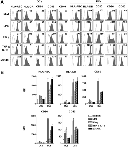
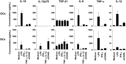
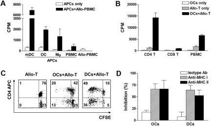
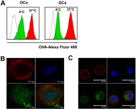
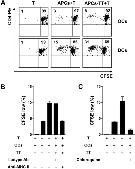
The bone and immune systems are closely related through cellular and molecular interactions. Because bone-resorbing osteoclasts (OCs) are derived from the monocyte/macrophage lineage, similar to dendritic cells (DCs), we hypothesized that OCs could serve as antigen-presenting cells (APCs) to activate T cells. In this study, OCs were generated from human monocytes with stimulation by receptor activator of nuclear factor kappaB ligand (RANKL) and macrophage colony-stimulating factor (M-CSF). Results showed that, similar to DCs, OCs express major histocompatibility complex (MHC) classes I and II, and CD80, CD86, and CD40; and uptake soluble antigens. OCs secrete interleukin-10 (IL-10), transforming growth factor-beta (TGF-beta), IL-6, and tumor necrosis factor-alpha (TNF-alpha), but not IL-12p70. OCs present allogeneic antigens and activate both CD4+ and CD8+ alloreactive T cells in an MHC-restricted fashion. OCs also present soluble protein tetanus toxoid to activate autologous CD4+ T cells. These findings indicate that OCs can function as APCs and activate both CD4+ and CD8+ T cells. Thus, our study provides new insight into the effect of OCs on the immune system and may help develop novel strategies for treating diseases such as rheumatoid arthritis and multiple myeloma, which affect both the bone and immune systems.


Figures

Figure 1
Figure 1
Characterization of OCs. Osteoclasts (OCs) were generated from monocytes with macrophage colony-stimulating factor (M-CSF) and receptor activator of nuclear factor κB ligand (RANKL) for 14 days. Dendritic cells (DCs) were derived from monocytes with the addition of granulocyte-macrophage colony-stimulating factor and IL-4 for 5 days. (A) Tartrate-resistant acid phosphatase (TRAP) staining to detect the TRAP activity in monocytes, OCs, and DCs. TRAP activity appears as purplish to dark red granules in the cytoplasm of the cells. (B) Bone resorption assay of OCs and DCs. OCs and DCs were generated from monocytes in 96-well plates with dentine discs in the wells. Dentine discs were stained with toluidine blue, and the blue area indicates the resorption of the dentine. (C) Phenotype of OCs and immature DCs. OCs and DCs were harvested and stained with antibodies and analyzed by flow cytometry. Shown are representative results of 3 independent experiments.
Figure 2
Figure 2
Phenotypic properties of OCs and DCs.(A) OCs and DCs were cultured with lipopolysaccharide (LPS), interferon-γ (IFN-γ), tumor necrosis factor-α (TNF-α), and interleukin-1β (IL-1β), or sCD40L for 48 hours. Cells were harvested and stained with antibodies for flow cytometric assay. (B) Summarized data (mean ± SEM) for mean fluorescence intensity (MFI) of cells from 3 different donors are shown.
Figure 3
Figure 3
Cytokine profiles of OCs and DCs. Monocyte-derived OCs and DCs were cultured with LPS, IFN-γ, TNF-α, and IL-1β, or sCD40L for 48 hours, supernatants were harvested, and cytokine concentration was measured by ELISA. Shown are summarized data (mean ± SEM) of cytokines from cells from 3 different donors.
Figure 4
Figure 4
Capacity of OCs to stimulate alloreactive T cells. (A) Allogeneic peripheral blood mononuclear cells (PBMCs; 2 × 10⁵) were cocultured with 104 LPS-matured DCs (mDCs), OCs, macrophages (Mφ), or PBMCs from the same donors (n = 3), and T-cell proliferation was determined on day 4 with 3H-thymidine assay. (B) OC capacity to activate both CD4+ and CD8+ alloreactive T cells in mixed lymphocyte reaction (MLR) assay. Allogeneic PBMCs or purified CD4+ or CD8+ T cells (2 × 10⁵) were cocultured with 104 OCs, and T-cell proliferation was determined on day 5. Summarized data (mean ± SEM) of cell proliferation (cpm) of cells from 3 different donors are shown. (C) OC capacity to activate both CD4+ and CD8+ alloreactive T cells measured by CFSE dilution assay. CFSE-labeled CD3+ T cells were cocultured with allogeneic OCs and DCs for 5 days. Representative results from 1 of 3 performed experiments are shown. (D) Major histocompatibility complex (MHC) restriction in MLR assay. Antibodies for HLA-ABC or HLA-DR (10 μg/mL) were added to the culture of MLR assay. Isotype IgG was used as control. Shown are percentages of inhibition (mean ± SD) from 3 independent experiments using cells from 3 different blood donors.
Figure 5
Figure 5
Cytokine production of allospecific CD4+ T cells. Purified CD3+ T cells were stimulated with allogeneic OCs or LPS-stimulated OCs for 6 days. DCs were used as control. After the culture, CD4+ T cells were sorted out and restimulated with anti-CD3 and anti-CD28 antibodies for 24 hours. Supernatants were collected and the concentrations of IFN-γ, IL-10, IL-4, and TGF-β1 were measured by ELISA. (A) Production of cytokines by CD4+ T cells stimulated by OCs. (B) Production of cytokines by CD4+ T cells stimulated by DCs. Representative results from 2 independent experiments are shown.
Figure 6
Figure 6
Uptake of soluble antigens by OCs. OCs and immature DCs were incubated with OVA–Alexa Fluor 488 at a concentration of 1 mg/mL at 37°C or 4°C for 30 minutes. After washing and fixation, cells were analyzed by flow cytometry and confocal microscope. (A) Flow cytometric analysis of the uptake of OVA–Alexa Fluor 488 by OCs and DCs. Unfilled curves show cells incubated without OVA–Alexa Fluor 488, green curves show cells incubated with OVA–Alexa Fluor 488 at 4°C, and red curves show cells incubated with OVA–Alexa Fluor 488 at 37°C. Images of (B) OCs or (C) DCs by confocal microscopy. Red shows staining with anti-CD11c antibody, green shows staining with OVA–Alexa Fluor 488, and blue shows nuclei stained with DAPI. Scale bar represents 20 μm. Representative results from 1 of 3 experiments performed are shown.
Figure 7
Figure 7
Capacity of OCs to present soluble antigen TT to CD4+ T cells and induce T-cell proliferation. (A) Proliferation of tetanus toxoid (TT)–specific T cells induced by TT-pulsed OCs and DCs. CFSE-labeled CD4+ T cells were cultured with autologous OCs or DCs pulsed with TT for 6 days. Flow cytometric analysis was performed to detect the proliferation of CD4+ T cells. Representative results from 1 of 3 experiments performed are shown. (B) MHC restriction in TT-specific CD4+ T-cell response. Antibody (10 μg/mL) for HLA-DR or isotypic control was added to the culture of T cells and TT-pulsed OCs. Data show the proliferation rate of CD4+ T cells detected by CFSE dilution assay. (C) Effect of chloroquine on antigen processing and presentation by OCs. Chloroquine was added at a final concentration of 50 mM to the cell cultures, and T-cell proliferation was detected by CFSE dilution assay. Cultures in panels B and C were run in triplicate and results are expressed as mean ± SD. Representative results from 2 independent experiments are shown.

References

    1. Katagiri T, Takahashi N. Regulatory mechanisms of osteoblast and osteoclast differentiation. Oral Dis. 2002;8(3):147–159. - PubMed
    1. Takayanagi H. Osteoimmunology: shared mechanisms and crosstalk between the immune and bone systems. Nat Rev Immunol. 2007;7(4):292–304. - PubMed
    1. Lorenzo J, Horowitz M, Choi Y. Osteoimmunology: interactions of the bone and immune system. Endocr Rev. 2008;29(4):403–440. - PMC - PubMed
    1. Wong BR, Rho J, Arron J, et al. TRANCE is a novel ligand of the tumor necrosis factor receptor family that activates c-Jun N-terminal kinase in T cells. J Biol Chem. 1997;272(40):25190–25194. - PubMed
    1. Wong BR, Josien R, Choi Y. TRANCE is a TNF family member that regulates dendritic cell and osteoclast function. J Leukoc Biol. 1999;65(6):715–724. - PubMed

Publication types

MeSH terms