

Weight loss in obese C57BL/6 mice limits adventitial expansion of established angiotensin II-induced abdominal aortic aneurysms
- PMID: 20304811
- PMCID: PMC2886622
- DOI: 10.1152/ajpheart.00961.2009
Weight loss in obese C57BL/6 mice limits adventitial expansion of established angiotensin II-induced abdominal aortic aneurysms
Abstract
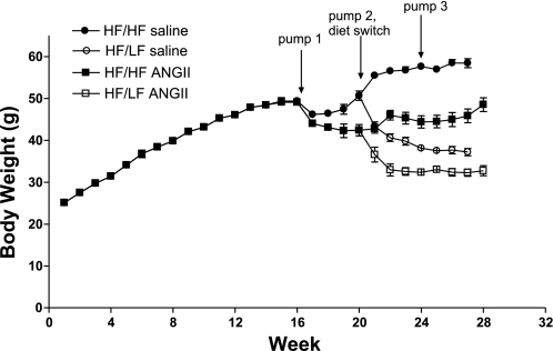
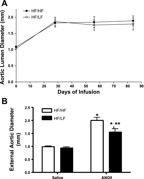
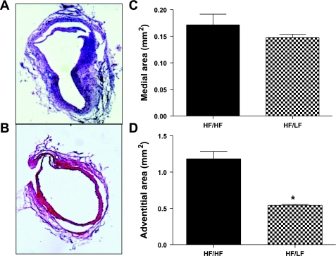
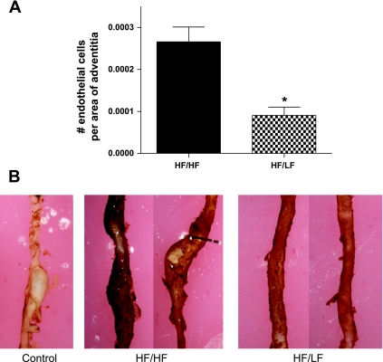
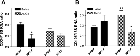
Previous studies demonstrated that obesity increases inflammation in periaortic adipose tissue and promotes angiotensin II (ANG II)-induced abdominal aortic aneurysms (AAAs). We sought to determine whether weight loss of obese C57BL/6 mice would influence the progression of established AAAs. Male C57BL/6 mice were fed a high-fat diet (HF) for 4 mo and then infused with either saline or ANG II (1,000 ng x kg(-1) x min(-1)) for 3 mo. Mice with dilated suprarenal aortas at 28 days of ANG II infusion were designated to groups fed the HF (HF/HF) or a low-fat diet (LF; 10% kcal as fat; HF/LF) to induce weight loss for the last 2 mo of infusions. Suprarenal aortic lumen diameters of obese mice were increased by ANG II infusion at day 28 (day 0: 1.03 + or - 0.02; day 28: 1.86 + or - 0.14 mm; P < 0.05), but did not progress with continued infusion in HF/HF mice. Moreover, aortic lumen diameters were not different between groups (HF/HF: 1.89 + or - 0.15; HF/LF: 1.79 + or - 0.18 mm). However, maximal diameters of excised AAAs were decreased with weight loss (HF/HF: 2.00 + or - 0.11; HF/LF: 1.55 + or - 0.13 mm; P < 0.05) and had reduced adventitial areas (HF/HF: 1.18 + or - 0.10; HF/LF: 0.54 + or - 0.02 mm(2); P < 0.05). Neovascularization of aortic adventitias was strikingly decreased in HF/LF mice (HF/HF: 43 + or - 5; HF/LF: 12 + or - 2 endothelial cells/adventitial area; P < 0.05). ANG II-induced elevations in adipose mRNA abundance of CD105, an adipose-derived stem cell marker, were abolished with weight loss. These results demonstrate that weight loss limits adventitial expansion of ANG II-induced AAAs. Reduced neovascularization from weight loss may limit progression of AAAs.
Figures
Similar articles
-
Obesity promotes inflammation in periaortic adipose tissue and angiotensin II-induced abdominal aortic aneurysm formation.Arterioscler Thromb Vasc Biol. 2009 Oct;29(10):1458-64. doi: 10.1161/ATVBAHA.109.192658. Epub 2009 Jul 16. Arterioscler Thromb Vasc Biol. 2009. PMID: 19608970 Free PMC article.
-
Castration of male mice prevents the progression of established angiotensin II-induced abdominal aortic aneurysms.J Vasc Surg. 2015 Mar;61(3):767-76. doi: 10.1016/j.jvs.2013.11.004. Epub 2014 Jan 16. J Vasc Surg. 2015. PMID: 24439319 Free PMC article.
-
Adipocyte-Derived Serum Amyloid A Promotes Angiotensin II-Induced Abdominal Aortic Aneurysms in Obese C57BL/6J Mice.Arterioscler Thromb Vasc Biol. 2022 May;42(5):632-643. doi: 10.1161/ATVBAHA.121.317225. Epub 2022 Mar 28. Arterioscler Thromb Vasc Biol. 2022. PMID: 35344382 Free PMC article.
-
Role of the renin-angiotensin system in the development of abdominal aortic aneurysms in animals and humans.Ann N Y Acad Sci. 2006 Nov;1085:82-91. doi: 10.1196/annals.1383.035. Ann N Y Acad Sci. 2006. PMID: 17182925 Review.
-
The role of the renin-angiotensin system in aortic aneurysmal diseases.Curr Hypertens Rep. 2008 Apr;10(2):99-106. doi: 10.1007/s11906-008-0020-3. Curr Hypertens Rep. 2008. PMID: 18474175 Free PMC article. Review.
Cited by
-
Limosilactobacillus reuteri HY7503 and Its Cellular Proteins Alleviate Endothelial Dysfunction by Increasing Nitric Oxide Production and Regulating Cell Adhesion Molecule Levels.Int J Mol Sci. 2024 Oct 21;25(20):11326. doi: 10.3390/ijms252011326. Int J Mol Sci. 2024. PMID: 39457107 Free PMC article.
-
Changes in aortic diameter induced by weight loss: The HELENA trial- whole-body MR imaging in a dietary intervention trial.Front Physiol. 2022 Sep 20;13:976949. doi: 10.3389/fphys.2022.976949. eCollection 2022. Front Physiol. 2022. PMID: 36203934 Free PMC article.
-
Quantitative Aortic Distensibility Measurement Using CT in Patients with Abdominal Aortic Aneurysm: Reproducibility and Clinical Relevance.Biomed Res Int. 2017;2017:5436927. doi: 10.1155/2017/5436927. Epub 2017 Apr 18. Biomed Res Int. 2017. PMID: 28484713 Free PMC article. Clinical Trial.
-
Aging exacerbates obesity-induced oxidative stress and inflammation in perivascular adipose tissue in mice: a paracrine mechanism contributing to vascular redox dysregulation and inflammation.J Gerontol A Biol Sci Med Sci. 2013 Jul;68(7):780-92. doi: 10.1093/gerona/gls238. Epub 2012 Dec 3. J Gerontol A Biol Sci Med Sci. 2013. PMID: 23213032 Free PMC article.
-
Perivascular adipose tissue-derived extracellular vesicle miR-221-3p mediates vascular remodeling.FASEB J. 2019 Nov;33(11):12704-12722. doi: 10.1096/fj.201901548R. Epub 2019 Aug 30. FASEB J. 2019. PMID: 31469602 Free PMC article.
References
-
- Barisione C, Charnigo R, Howatt DA, Moorleghen JJ, Rateri DL, Daugherty A. Rapid dilation of the abdominal aorta during infusion of angiotensin II detected by noninvasive high-frequency ultrasonography. J Vasc Surg 44: 372–376, 2006 - PubMed
-
- Bentzon JF, Sondergaard CS, Kassem M, Falk E. Smooth muscle cells healing atherosclerotic plaque disruptions are of local, not blood, origin in apolipoprotein E knockout mice. Circulation 116: 2053–2061, 2007 - PubMed
-
- Cancello R, Henegar C, Viguerie N, Taleb S, Poitou C, Rouault C, Coupaye M, Pelloux V, Hugol D, Bouillot JL, Bouloumié A, Barbatelli G, Cinti S, Svensson PA, Barsh GS, Zucker JD, Basdevant A, Langin D, Clément K. Reduction of macrophage infiltration and chemoattractant gene expression changes in white adipose tissue of morbidly obese subjects after surgery-induced weight loss. Diabetes 54: 2277–2286, 2005 - PubMed
-
- Capel F, Klimcakova E, Viguerie N, Roussel B, Vítková M, Kováciková M, Polák J, Kovácová Z, Galitzky J, Maoret JJ, Hanácek J, Pers TH, Bouloumié A, Stich V, Langin D. Macrophages and adipocytes in human obesity: adipose tissue gene expression and insulin sensitivity during calorie restriction and weight stabilization. Diabetes 58: 1558–1567, 2009 - PMC - PubMed
Publication types
MeSH terms
Substances
Grants and funding
LinkOut - more resources
Full Text Sources
Medical
Research Materials
Miscellaneous