. 2010 May 14;285(20):15201-15208.
doi: 10.1074/jbc.M110.101808. Epub 2010 Mar 19.

Transcription-dependent activation of ataxia telangiectasia mutated prevents DNA-dependent protein kinase-mediated cell death in response to topoisomerase I poison

Affiliations

Transcription-dependent activation of ataxia telangiectasia mutated prevents DNA-dependent protein kinase-mediated cell death in response to topoisomerase I poison

Ryo Sakasai et al. J Biol Chem. .

Abstract

Camptothecin (CPT) is a topoisomerase I inhibitor, derivatives of which are being used for cancer chemotherapy. CPT-induced DNA double-strand breaks (DSBs) are considered a major cause of its tumoricidal activity, and it has been shown that CPT induces DNA damage signaling through the phosphatidylinositol 3-kinase-related kinases, including ATM (ataxia telangiectasia mutated), ATR (ATM and Rad3-related), and DNA-PK (DNA-dependent protein kinase). In addition, CPT causes DNA strand breaks mediated by transcription, although the downstream signaling events are less well characterized. In this study, we show that CPT-induced activation of ATM requires transcription. Mechanistically, transcription inhibition suppressed CPT-dependent activation of ATM and blocked recruitment of the DNA damage mediator p53-binding protein 1 (53BP1) to DNA damage sites, whereas ATM inhibition abrogated CPT-induced G(1)/S and S phase checkpoints. Functional inactivation of ATM resulted in DNA replication-dependent hyperactivation of DNA-PK in CPT-treated cells and dramatic CPT hypersensitivity. On the other hand, simultaneous inhibition of ATM and DNA-PK partially restored CPT resistance, suggesting that activation of DNA-PK is proapoptotic in the absence of ATM. Correspondingly, comet assay and cell cycle synchronization experiments suggested that transcription collapse occurring as the result of CPT treatment are converted to frank double-strand breaks when ATM-deficient cells bypass the G(1)/S checkpoint. Thus, ATM suppresses DNA-PK-dependent cell death in response to topoisomerase poisons, a finding with potential clinical implications.


Figures
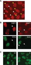
FIGURE 1.
FIGURE 1.
CPT induces two distinct types of 53BP1 foci. A, representative image of 53BP1 foci induced by CPT (2 μm, 1 h) treatment. The HeLa cells denoted by white arrowheads represent Type I 53BP1 foci, whereas yellow arrowheads denote Type II foci. B, Type I 53BP1 foci occurring in non-S phase cells. HeLa cells were treated with CPT (2 μm, 1 h) and stained with anti-53BP1 and RPA2 antibodies (left). To observe DNA synthesis, cells were treated with BrdUrd (20 μm, 20 min) and stained with α-53BP1 and α-BrdUrd antibodies (right). C, CPT-induced 53BP1 foci in G1 or S phase cells. Cells were synchronized in G1 phase and S phase by release from nocodazole block and thymidine block, respectively. After treatment with CPT (2 μm, 1 h), cells were stained with anti-53BP1 antibody.
FIGURE 2.
FIGURE 2.
Type I 53BP1 foci are ATM-dependent and occur in response to transcription-mediated DNA damage. A, co-staining 53BP1 with γH2AX. HeLa cells were treated with CPT (2 μm, 1 h) and stained with α-53BP1 and γH2AX antibodies. B, formation of 53BP1 foci requiring RNF8. HeLa cells were transfected with nontargeting (siCTR) or RNF8-targeting siRNA (siRNF8) and stained with anti-53BP1 antibody following CPT (2 μm, 1 h) treatment. C, effect of ATM inhibition on Type I 53BP1 foci formation. HeLa cells were treated with solvent only, KU-55933 (10 μm, 1 h) before CPT (2 μm, 1 h) treatment, and stained with anti-53BP1 and anti-RPA2 antibodies. D, Type I 53BP1 foci in ATM-deficient cells. Control cells (SuSa/Tn) and ATM-deficient cells (AT1OS/Tn) were treated with CPT (2 μm, 1 h) and stained with anti-53BP1 and anti-RPA2 antibodies. E, effect of transcription inhibition on Type I 53BP1 foci formation. HeLa cells were treated with DRB (100 μm, 2 h) before CPT (2 μm, 1 h) treatment and stained with anti-53BP1 and anti-RPA2 antibodies. The number of 53BP1 foci observed in cells without RPA2 signal was counted. Error bars show S.E. calculated from three independent experiments.
FIGURE 3.
FIGURE 3.
Transcription-dependent ATM activation in response to CPT. HeLa cells were pretreated with DNA replication inhibitors (HU and thymidine (Thy), 2 mm and 2.5 mm, respectively) or transcription inhibitors (DRB and α-amanitin, 100 μm and 5 μm, respectively) before CPT (2 μm, 1 h) treatment. DNA damage signaling was analyzed by Western blotting using appropriate antibodies.
FIGURE 4.
FIGURE 4.
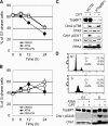
CPT induces ATM-dependent G1/S and S phase checkpoint arrest. A and B, U2OS cells were treated with 1 μm CPT following treatment with ATM (KU-55933; 10 μm, 1 h) or DNA-PK inhibitor (NU7026; 10 μm, 1 h). After incubation for the indicated time, cells were fixed with ethanol for propidium iodide staining. Cell cycle distribution was analyzed by flow cytometry, and the percentage of cells in G1 phase and S phase was plotted in A and B, respectively. Error bars show S.E. calculated from three independent experiments. C, CPT-induced Chk1 phosphorylation is TopBP1-dependent. HeLa cells transfected with control (siCTR) or TopBP1 siRNA (siTopBP1) were treated with CPT (2 μm, 1 h), and then Chk2, Chk1, and RPA2 phosphorylation were analyzed by Western blotting with the indicated antibodies. D, CPT-induced TopBP1 phosphorylation is restricted to S phase. To synchronize cell cycle in the G1 and S phase, HeLa cells were released from nocodazole block and thymidine block, respectively. Both cell populations were treated with CPT (2 μm, 1 h) with or without ATM inhibitor (KU-55933; 10 μm, 1 h). TopBP1, Chk1, and Chk1 phosphorylation was detected by Western blotting using the indicated antibodies.
FIGURE 5.
FIGURE 5.
ATM suppresses DNA-PK activation in response to CPT. A, effects of ATM, replication, and transcription inhibitors on CPT-induced DNA-PKcs autophosphorylation. HeLa cells were treated with CPT (2 μm, 1 h) following ATM inhibitor (KU-55933; 10 μm, 1 h) with HU (2 mm, 10 min) or DRB (100 μm, 2 h) treatment, and DNA-PKcs autophosphorylation and Chk2 phosphorylation were analyzed by Western blotting. B, DNA-PK activation in ATM-deficient cells. Control cells (GM00637H or SuSa/Tn) and ATM-deficient cells (GM05849C or AT1OS/Tn) were treated with CPT (2 μm, 1 h), and DNA-PKcs autophosphorylation were analyzed by Western blotting. C, comet assay for detection of CPT-induced DNA strand breaks. HeLa cells were treated with CPT (0.25 μm) for 1 h following ATM inhibitor (10 μm, 1 h) treatment in the absence or presence of DRB (100 μm, 2 h), and induced-DNA strand breaks were detected by neutral comet assay. The comet tail moments were averaged in triplicate experiments, where the median among 100 cells was calculated in each experiment. Error bars represent S.E. calculated from three independent experiments. D, inhibition of ATM leads to DNA damage carry over from G1 to S phase. U2OS cells were synchronized in M phase with nocodazole and released into G1 and S phase upon incubation with fresh medium. Flow cytometry histograms show cell cycle profiles after release from the nocodazole block in the absence or presence of KU-55933 (10 μm, 1 h). Where indicated, cells were treated with CPT (5 μm, 1 h). The status of DNA-PK activation in the different experimental samples was determined by Western blotting with anti-phospho-DNA-PK antibodies.
FIGURE 6.
FIGURE 6.
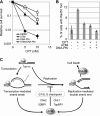
DNA-PK activation promotes cells death in response to CPT. A, clonogenic survival assays of HCT-116 cells treated with CPT in the absence or presence of ATM and/or DNA-PK inhibitors. Cells pretreated with ATM inhibitor (KU-55933; 10 μm, 1 h) and/or DNA-PK inhibitor (NU7026; 10 μm, 1 h) were cultured in the presence of CPT for 2 days at the indicated concentration. Error bars show S.E. calculated from three independent experiments. B, effects of ATM and/or DNA-PK inhibition on Rad51 foci formation. HeLa cells were treated with CPT (2 μm, 6 h) following pretreatment with ATM (10 μm, 1 h) and/or DNA-PK (10 μm, 1 h) inhibitors, and then cells were stained with anti-Rad51 antibody. The cells with over 10 foci were considered Rad51 foci-positive, and the percentage of Rad51 foci-positive cells is depicted graphically. Error bars show S.E. calculated from three independent experiments. C, schematic model summarizing the CPT-induced cellular responses. Transcription-mediated strand breaks caused by CPT in G1 phase are converted to double strand end by DNA replication. ATM is activated against both DNA damages and induces G1/S and S phase checkpoints to prevent DNA-PK hyperactivation leading to cell death.

Similar articles

Cited by

References

    1. Pommier Y. (2006) Nat. Rev. Cancer 6, 789–802 - PubMed
    1. Arnaudeau C., Lundin C., Helleday T. (2001) J. Mol. Biol. 307, 1235–1245 - PubMed
    1. Klein H. L., Kreuzer K. N. (2002) Mol. Cell 9, 471–480 - PubMed
    1. Cliby W. A., Lewis K. A., Lilly K. K., Kaufmann S. H. (2002) J. Biol. Chem. 277, 1599–1606 - PubMed
    1. Wang J. L., Wang X., Wang H., Iliakis G., Wang Y. (2002) Cell Cycle 1, 267–272 - PubMed

Publication types

MeSH terms