. 2010 May 14;285(20):14999-15009.
doi: 10.1074/jbc.M110.109819. Epub 2010 Mar 19.

TAX1BP1 and A20 inhibit antiviral signaling by targeting TBK1-IKKi kinases

Affiliations

TAX1BP1 and A20 inhibit antiviral signaling by targeting TBK1-IKKi kinases

Kislay Parvatiyar et al. J Biol Chem. .

Abstract

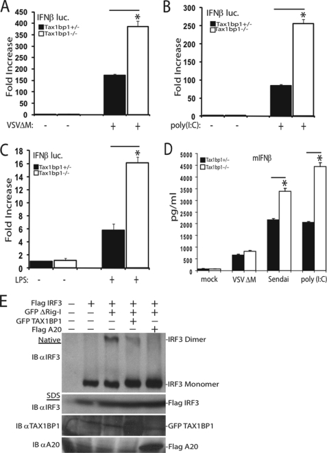
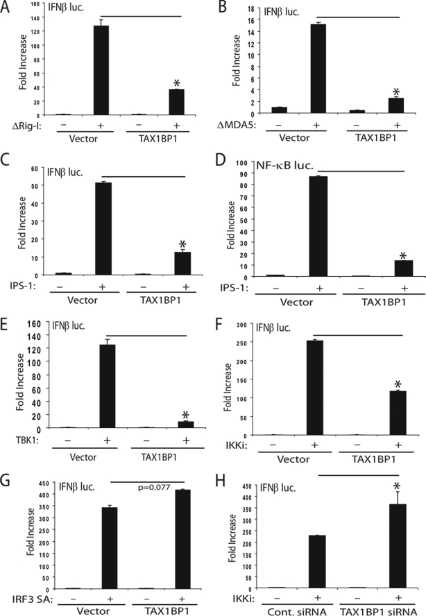
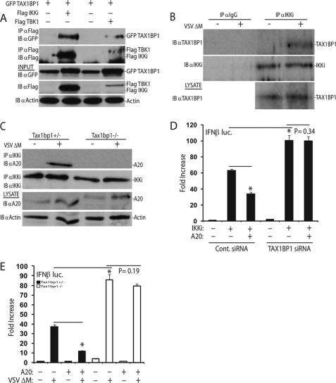
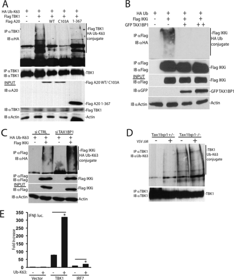
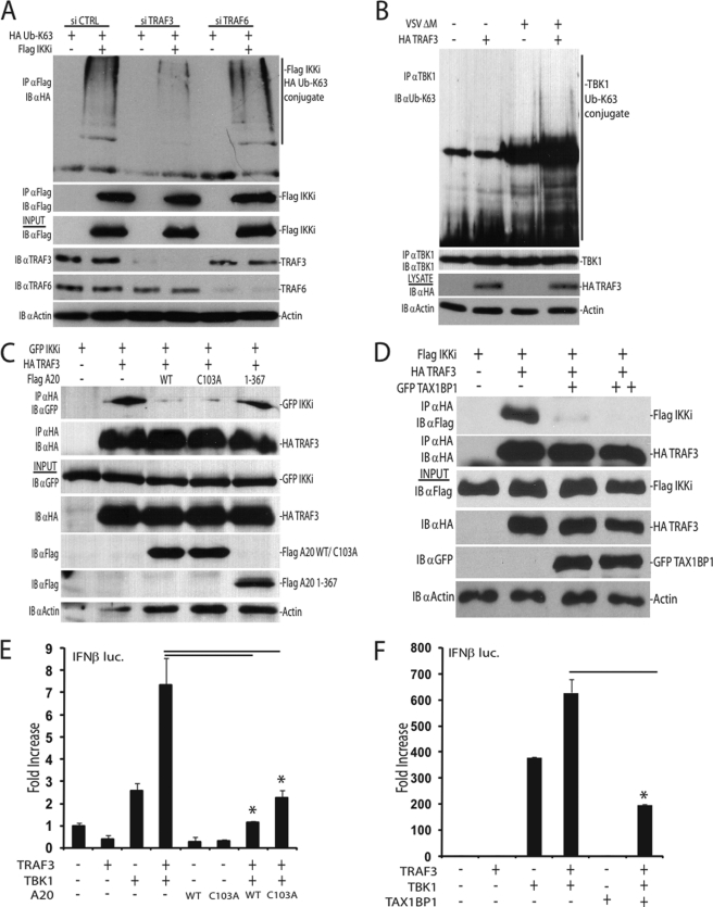
Induction of type I interferons by the transcription factor IRF3 is essential in the initiation of antiviral innate immunity. Activation of IRF3 requires C-terminal phosphorylation by the upstream kinases TBK1-IKKi, where IRF3 phosphorylation promotes dimerization, and subsequent nuclear translocation to the IFNbeta promoter. Recent studies have described the ubiquitin-editing enzyme A20 as a negative regulator of IRF3 signaling by associating with TBK1-IKKi; however, the regulatory mechanism of A20 inhibition remains unclear. Here we describe the adaptor protein, TAX1BP1, as a key regulator of A20 function in terminating signaling to IRF3. Murine embryonic fibroblasts (MEFs) deficient in TAX1BP1 displayed increased amounts of IFNbeta production upon viral challenge compared with WT MEFs. TAX1BP1 inhibited virus-mediated activation of IRF3 at the level of TBK1-IKKi. TAX1BP1 and A20 blocked antiviral signaling by disrupting Lys(63)-linked polyubiquitination of TBK1-IKKi independently of the A20 deubiquitination domain. Furthermore, TAX1BP1 was required for A20 effector function because A20 was defective for the targeting and inactivation of TBK1 and IKKi in Tax1bp1(-)(/)(-) MEFs. Additionally, we found the E3 ubiquitin ligase TRAF3 to play a critical role in promoting TBK1-IKKi ubiquitination. Collectively, our results demonstrate TBK1-IKKi to be novel substrates for A20 and further identify a novel mechanism whereby A20 and TAX1BP1 restrict antiviral signaling by disrupting a TRAF3-TBK1-IKKi signaling complex.


Figures

FIGURE 1.
FIGURE 1.
TAX1BP1 is a negative regulator of antiviral signaling. A–C, Tax1bp1+/− or Tax1bp1−/− MEFs were transfected with an IFNβ luc reporter (200 ng) and pRL-tk (20 ng). Cells were virally infected (A), treated with poly(I-C) (25 μg/ml) (B), or treated with lipopolysaccharide (1 μg/ml) (C). IFNβ luciferase assays were performed 16 h after the respective stimulations. D, Tax1bp1+/− or Tax1bp1−/− MEFs were mock-infected, virally infected, or transfected with poly(I-C) (6 μg) for 16 h. Supernatants were subjected to IFNβ ELISA. E, 293T cells were transfected with the indicated plasmids (1 μg of FLAG-IRF3, GFP-TAX1BP1, or FLAG-A20 and 100 ng of GFP-ΔRig-I), and lysates were subjected to native PAGE (top) or SDS-PAGE (bottom) and immunoblotted (IB) with the indicated antibodies. Error bars, S.E.
FIGURE 2.
FIGURE 2.
TAX1BP1 inhibits IFNβ activation at the level of TBK1-IKKi. A–C, 293T cells were transfected with IFNβ luc reporter (100 ng) and pRL-tk (10 ng) plasmids together with ΔRig-I (A), ΔMDA5 (B), or IPS-1 (C) (1 μg) and a plasmid expressing TAX1BP1 (1 μg). D, 293T cells were transfected as described in C except an NF-κB luc reporter (100 ng) was used. E–G, 293T cells were transfected as in A–C using plasmids (1 μg each) encoding either TBK1 (E), IKKi (F), or a constitutively active form of IRF3 (IRF3 SA) (G) with or without TAX1BP1 DNA (1 μg). H, 293T cells were transfected with either control (Cont.) scrambled or TAX1BP1 siRNA. After 24 h, cells were transfected with IFNβ luc, pRL-tk, and IKKi plasmids as in F. Error bars, S.E.
FIGURE 3.
FIGURE 3.
A20 requires TAX1BP1 to terminate antiviral signaling. A, 293T cells were transfected with the indicated plasmids (1 μg each). Co-IP and immunoblots were performed with the indicated antibodies. B, IKKi was immunoprecipitated from whole cell lysates derived from mock-infected and virally infected wild-type MEFs. Bound proteins were detected via immunoblotting, and lysates were probed as indicated. C, Tax1bp1+/− or Tax1bp1−/− MEFs were mock-infected or virally infected for 16 h, and lysates were subjected to immunoprecipitation and/or immunoblotting using the indicated antibodies. D, 293T cells were transfected with either control scrambled siRNA or TAX1BP1 siRNA. After 24 h, cells were transfected with IFNβ luc (100 ng) and pRL-tk (10 ng) DNA and plasmids encoding IKKi (1 μg) and A20 (200 ng) as indicated. E, Tax1bp1+/− or Tax1bp1−/− MEFs were transfected with an IFNβ luc reporter (200 ng) and pRL-tk (20 ng) and a plasmid encoding A20 (1 μg) as indicated. After 36 h, cells were either mock-infected or infected with virus for 16 h, followed by luciferase assays. IB, immunoblot; IP, immunoprecipitation. Error bars, S.E.
FIGURE 4.
FIGURE 4.
TAX1BP1 and A20 target TBK1-IKKi for deubiquitination. A, 293T cells were cotransfected with epitope-tagged constructs as indicated (1 μg of FLAG-TBK1, FLAG-A20 WT, FLAG-A20 C103A, and FLAG A20 1–367 and 500 ng of HA-Ub Lys63-only). Co-IPs and immunoblots evaluating TBK1 ubiquitination and immunoprecipitation were performed using the indicated antibodies. B, epitope-tagged constructs (1 μg of FLAG-IKKi, 500 ng of HA-Ub, and 1 or 2 μg of GFP-TAX1BP1) were transfected into 293T cells. Co-IPs and immunoblots evaluating IKKi ubiquitination were performed as in A. C, 293T cells were transfected with control scrambled siRNA or siRNA targeting TAX1BP1. After 24 h, cells were cotransfected with the indicated plasmids (1 μg of FLAG-IKKi and 500 ng of HA-Ub Lys63-only), and IKKi polyubiquitination was determined as in B. D, Tax1bp1+/− or Tax1bp1−/− MEFs were mock-infected or virally infected for 16 h, and lysates were subjected to immunoprecipitation and/or immunoblotting as indicated. E, 293T cells were cotransfected with IFNβ luc (100 ng) and pRL-tk (10 ng) DNA and plasmids encoding TBK1 or IRF7 (1 μg) and Lys63-only ubiquitin (500 ng) as indicated. WT, wild type; IB, immunoblot; IP, immunoprecipitation.
FIGURE 5.
FIGURE 5.
A20 and TAX1BP1 disrupt a TRAF3-TBK1-IKKi signaling module. A, 293T cells were transfected with either control scrambled siRNA, TRAF3 siRNA, or TRAF6 siRNA. After 24 h, cells were transfected with plasmids as indicated (1 μg of FLAG-IKKi, 500 ng of HA-Ub Lys63-only). Cell lysates were subjected to immunoprecipitation and immunoblotting with the indicated antibodies. B, TRAF3-deficient MEFs were transfected with either empty vector DNA or a plasmid expressing TRAF3 (2.5 μg). After 36 h, MEFs were mock-infected or virally infected for 16 h. Lysates were subjected to immunoprecipitation and/or immunoblotting with the indicated antibodies. C and D, A20 and TAX1BP1 disrupt TRAF3-IKKi interactions. C, 293T cells were transfected with the indicated plasmids (1 μg of GFP-IKKi, HA-TRAF3, FLAG-A20 WT, FLAG-A20 C103A, or FLAG-A20 1–367). Whole cell lysates were subjected to co-IPs and/or immunoblotting with the indicated antibodies. D, 293T cells were cotransfected with HA-TRAF3 and FLAG-IKKi (1 μg each) and either 1 or 2 μg of GFP-TAX1BP1 plasmid DNAs as indicated. Lysates were subjected to co-IPs and immunoblotting as in C. E and F, 293T cells were transfected with IFNβ luc (100 ng) and pRL-tk (10 ng) DNA together with the indicated plasmids (1 μg each). WT, wild type; IB, immunoblot; IP, immunoprecipitation. Error bars, S.E.

References

    1. Kawai T., Akira S. (2008) Ann. N.Y. Acad. Sci. 1143, 1–20 - PubMed
    1. Hiscott J., Grandvaux N., Sharma S., Tenoever B. R., Servant M. J., Lin R. (2003) Ann. N.Y. Acad. Sci. 1010, 237–248 - PubMed
    1. Sadler A. J., Williams B. R. (2008) Nat. Rev. Immunol. 8, 559–568 - PMC - PubMed
    1. Medzhitov R. (2007) Nature 449, 819–826 - PubMed
    1. Honda K., Taniguchi T. (2006) Nat. Rev. Immunol. 6, 644–658 - PubMed

Publication types

MeSH terms