. 2010 May 14;285(20):15314-15323.
doi: 10.1074/jbc.M110.104281. Epub 2010 Mar 19.

The signal peptide of the IgE receptor alpha-chain prevents surface expression of an immunoreceptor tyrosine-based activation motif-free receptor pool

Affiliations

The signal peptide of the IgE receptor alpha-chain prevents surface expression of an immunoreceptor tyrosine-based activation motif-free receptor pool

Barbara Platzer et al. J Biol Chem. .

Abstract

The high affinity receptor for IgE, Fc epsilon receptor I (FcepsilonRI), is an activating immune receptor and key regulator of allergy. Antigen-mediated cross-linking of IgE-loaded FcepsilonRI alpha-chains induces cell activation via immunoreceptor tyrosine-based activation motifs in associated signaling subunits, such as FcepsilonRI gamma-chains. Here we show that the human FcepsilonRI alpha-chain can efficiently reach the cell surface by itself as an IgE-binding receptor in the absence of associated signaling subunits when the endogenous signal peptide is swapped for that of murine major histocompatibility complex class-I H2-K(b). This single-chain isoform of FcepsilonRI exited the endoplasmic reticulum (ER), trafficked to the Golgi and, subsequently, trafficked to the cell surface. Mutational analysis showed that the signal peptide regulates surface expression in concert with other described ER retention signals of FcepsilonRI-alpha. Once the FcepsilonRI alpha-chain reached the cell surface by itself, it formed a ligand-binding receptor that stabilized upon IgE contact. Independently of the FcepsilonRI gamma-chain, this single-chain FcepsilonRI was internalized after receptor cross-linking and trafficked into a LAMP-1-positive lysosomal compartment like multimeric FcepsilonRI. These data suggest that the single-chain isoform is capable of shuttling IgE-antigen complexes into antigen loading compartments, which plays an important physiologic role in the initiation of immune responses toward allergens. We propose that, in addition to cytosolic and transmembrane ER retention signals, the FcepsilonRI alpha-chain signal peptide contains a negative regulatory signal that prevents expression of an immunoreceptor tyrosine-based activation motif-free IgE receptor pool, which would fail to induce cell activation.


Figures
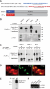
FIGURE 1.
FIGURE 1.
Properly folded FcϵRI-α reaches the Golgi in the absence of FcϵRI-γ. A, signal peptide (SP) sequences of FcϵRI-α (NCBI RefSeq NP_001992.1) and mouse major histocompatibility complex class I H2-Kb (NCBI RefSeq NP001001892.2) are depicted. The stretch of hydrophobic amino acids representing the predicted transmembrane region of the signal peptide is underlined. The construct containing the wild type FcϵRI-α chain with its endogenous signal peptide is referred to as endo-α, and the chimeric construct where the endogenous signal peptide was swapped for the signal peptide of H2-Kb is referred to as Kb-α. B, FcϵRI α-chain shows a protein pattern characteristic for ER- and Golgi-modified forms in the absence of γ-chain. 293T cells were transfected with indicated HA-tagged α-chain constructs or cotransfected with γ-chain as control (first lane 1). Smaller quantities of α-chain from endo-α transfected cells reached the Golgi compared with Kb-α transfected cells (second and third lanes). A construct without signal peptide (no SP) did not enter the ER (fourth lane). Immunoblot analysis was performed under nonreducing conditions with the anti-HA antibody 3F10 (upper blot). The blot was stripped and reprobed with a polyclonal anti-γ-chain reagent (lower blot). C, glycosylation study performed on the FcϵRI α-chain after immunoprecipitation with IgE. FcϵRI α-chain (first, fourth, and seventh lanes) is compared for its susceptibility to EndoH (second, fifth, and eighth lanes) or PNGase F (third, sixth, and ninth lanes) digestion. D, visualization of FcϵRI-γ independent trafficking to the Golgi. Immunofluorescence staining of MelJuso cells transfected with Kb-α. The FcϵRI α-chain was detected with the polyclonal rabbit serum 997 and anti-rabbit Alexa Fluor 568 (left images, shown in red). The Golgi was visualized with anti-GM130 followed by anti-mouse Alexa Fluor 488 (middle image, shown in green). The merged images are depicted in the right picture. E, molecular characteristics of α-chain from MelJuso cells transfected with Kb-α. Immunoprecipitation was performed with IgE; IgG was used to control for specificity of IgE binding. F, RT-PCR analysis confirmed lack of γ-chain expression in 239T, HeLa, and MelJuso cells. cDNA from human tonsil tissue was used as positive control (CTRL).
FIGURE 2.
FIGURE 2.
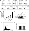
Cell surface expression of FcϵRI-α is regulated by its signal peptide. A, 293T, HeLa, and MelJuso cells were transfected with endo-α or Kb-α in vector pcDNA3.1 in the absence of the γ-chain. Cell surface expression of the α-chain was determined by flow cytometry. The gate depicting percentages of cells expressing α-chain at their cell surface was set accordingly to empty vector transfectants. B, eGFP expression correlates with surface expression of FcϵRI-α. 293T cells were transfected with Kb-α in pIRES2-eGFP. In the upper FACS dot plot eGFP expression is depicted. Three different regions were set corresponding to eGFPnegative (R1), eGFPlow (R2), and eGFPhigh (R3) cells. In the lower dot plots, α-chain expression at the cell surface was analyzed separately for these three different regions. C, 293T cells were transfected with endo-α and Kb-α constructs in pIRES2-eGFP. Empty vector transfected cells (CTRL) or cells cotransfected with Kb-α plus γ-chains were used as controls. The cells expressing equal intensities of eGFP were FACS-sorted, and cell lysates were analyzed for the protein characteristics of FcϵRI-α by immunoblotting with the mAb 19-1. Detection of eGFP with a polyclonal anti-GFP serum was used as a loading control. D, FcϵRI-α mRNA of cells that were FACS-sorted for equal levels of eGFP expression was determined by quantitative RT-PCR. E, the H2-Kb signal peptide drives surface expression of FcϵRI-α more efficiently than the endogenous signal peptide. Endo-α and Kb-α transfectants were gated based on equal eGFP expression and analyzed for surface α-chain. Empty pIRES2-eGFP vector transfected cells (CTRL) were used as control. The bar diagram represents the means ± S.E. of seven independent experiments.
FIGURE 3.
FIGURE 3.
The signal peptide regulates cell surface expression of the FcϵRI α-chain independently of ER retention signals in the cytosolic tail. A, 293T cells were transfected with constructs lacking the cytosolic tail of the α-chain (i.e. endo-αtail-minus and Kbtail-minus) or with the full-length constructs, endo-α and Kb-α, in pIRES2-eGFP. Transfected cells expressing equal levels of eGFP were gated (upper dot plots) and analyzed for α-chain at the cell surface (lower dot plots). Empty vector transfected cells were used as control (CTRL). B, quantification (means ± S.E.) of cell surface expression of 4 independent experiments as shown in A. C, cells expressing equal levels of eGFP were FACS-sorted and cell lysates were analyzed for protein characteristics of FcϵRI-α by immunoblotting with mAb 19-1. Detection of eGFP was used as loading control. D, an Asp → Asn mutation in the transmembrane domain increases surface expression of FcϵRI α-chain. HeLa cells were transiently transfected with Kb-α or the Asp → Asn transmembrane mutant (i.e. KbD/N). FACS histogram overlay depicts surface α-chain expression. Kb-α, filled, gray histogram; Kb-α D/N, striped, black histogram; empty vector, light gray line. E, relative FcϵRI-α mRNA levels of Kb-α and Kb-α D/N were determined by real time PCR. The values were determined in triplicate and normalized to glyceraldehyde-3-phosphate dehydrogenase expression. Representative data of three independent experiments are shown in D and E.
FIGURE 4.
FIGURE 4.
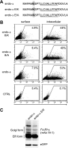
Point mutations in the endogenous signal peptide do not change cell surface transport of FcϵRI α-chain. A, scheme of introduced point mutations at position 6 of the endogenous signal peptide. B, 293T cells were transfected with endo-α, endo-α E/K, endo-α E/A, or empty vector. The data are representative of three independent experiments. C, cells expressing equal intensities of eGFP were FACS-sorted. The cell lysates were analyzed for the protein characteristics of FcϵRI-α by immunoblotting with mAb 19-1. Detection of eGFP was used as loading control.
FIGURE 5.
FIGURE 5.
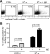
IgE-induced stabilization of FcϵRI at the cell surface does not require FcϵRI-γ-chains. A, 293T cells were transfected with Kb-α in pIRES2-eGFP, incubated with 200 ng/ml IgE for 18h, and analyzed by flow cytometry. Transfected cells were identified by eGFP expression and gated to determine cell surface expression of the α-chain. B, IgE stabilization of the single FcϵRI α-chain is signal peptide-independent. 293T cells were transfected with endo-α or Kb-α in pIRES2-eGFP, incubated with 200 ng/ml IgE for 18 h before analysis by flow cytometry. The data are presented as the means ± S.E. of three independent experiments. CTRL, control.
FIGURE 6.
FIGURE 6.
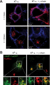
Single FcϵRI α-chain internalizes after receptor cross-linking. A, Kb-α was transiently transfected into HeLa cells (left images) or into HeLa cells stably expressing the FcϵRI γ-chain (right images). For FcϵRI-α cross-linking, the cells were incubated with the mouse anti-human FcϵRI-α antibody, CRAI, which was next cross-linked with an anti-mouse Alexa-Fluor 568-F(ab′)2 fragment (shown in red). The cells were incubated for 30 min at 37 °C (lower panels). For time point (t) = 0, the cells were fixed before cross-linking with anti-mouse Alexa Fluor 568-F(ab′)2 (upper panels). Cell surface membranes were visualized using Alexa Fluor 647-labeled wheat germ agglutinin (shown in blue). Confocal micrographs are presented as overlays. Representative experiment is shown (n = 3). B, FcϵRI α-chain traffics into LAMP-1 positive lysosomal compartments independently of the presence of FcϵRI-γ. HeLa cells expressing LAMP-1-eGFP (shown in green) were transfected with Kb-α (shown in red). Receptor cross-linking and internalization was performed as described above. The left panel shows cells transfected with Kb-α alone. The right panel shows cells that co-express FcϵRI α- and γ-chains. The bottom panel depicts higher magnifications of the lysosomal regions of the images shown in the upper panel. The bottom panel shows single channel images for LAMP-eGFP in green (left), FcϵRI-α in red (middle), and the merged image (right). The cells were analyzed 90 min after cross-linking.
FIGURE 7.
FIGURE 7.
Schematic of intracellular trafficking of FcϵRI and receptor-mediated cell activation. A, the subunits of multimeric FcϵRI complexes assemble in the ER, traffic to the Golgi and reach the cell surface. Antigen-induced cross-linking of IgE-loaded FcϵRI complexes induces ITAM-mediated cell activation and internalization of the receptor. Cross-linked FcϵRI shuttles antigen-IgE immune complexes to lysosomal compartments. B, single FcϵRI α-chain can exit the ER, traffic to the Golgi and reach the cell surface by itself. Trafficking of this single receptor isoform is tightly regulated by the signal peptide. Antigen-induced cross-linking of IgE-loaded single FcϵRI α-chain induces receptor internalization in the absence of ITAM-mediated cell activation. Like multimeric FcϵRI complexes, cross-linked single FcϵRI α-chain isoforms reach lysosomal compartments.

Similar articles

Cited by

References

    1. Abramson J., Pecht I. (2007) Immunol. Rev. 217, 231–254 - PubMed
    1. Gould H. J., Sutton B. J. (2008) Nat. Rev. Immunol. 8, 205–217 - PubMed
    1. Kraft S., Kinet J. P. (2007) Nat. Rev. Immunol. 7, 365–378 - PubMed
    1. Call M. E., Wucherpfennig K. W. (2005) Annu. Rev. Immunol. 23, 101–125 - PubMed
    1. Call M. E., Wucherpfennig K. W. (2007) Nat. Rev. Immunol. 7, 841–850 - PubMed

Publication types