. 2010 Jun;333(3):726-35.
doi: 10.1124/jpet.110.165837. Epub 2010 Mar 19.

A functional role for nicotinic acid adenine dinucleotide phosphate in oxytocin-mediated contraction of uterine smooth muscle from rat

Affiliations

A functional role for nicotinic acid adenine dinucleotide phosphate in oxytocin-mediated contraction of uterine smooth muscle from rat

Parvinder K Aley et al. J Pharmacol Exp Ther. 2010 Jun.

Abstract

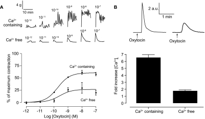
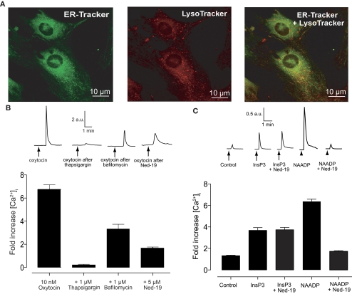
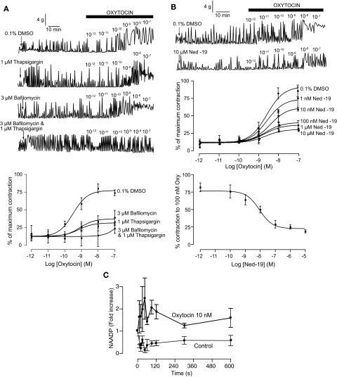
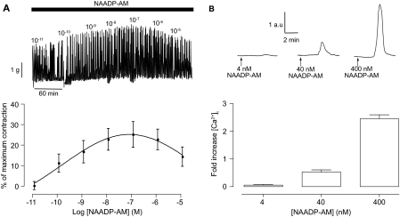
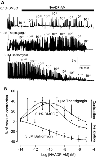
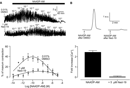
Conventionally, G protein-coupled receptors are thought to increase calcium via inositol 1,4,5-trisphosphate (InsP(3)). More recent evidence shows that an alternative second messenger, nicotinic acid adenine dinucleotide phosphate (NAADP), also has a role to play, causing researchers to question established calcium releasing pathways. With the recent development, by our group, of cell-permeant NAADP (NAADP-aceteoxymethyl ester) and a selective NAADP receptor antagonist (Ned-19; 1-(3-((4-(2-fluorophenyl)piperazin-1-yl)methyl)-4-methoxyphenyl)-2,3,4,9-tetrahydro-1H-pyrido[3,4-b]indole-3-carboxylic acid),the ability to investigate this signaling pathway has improved. Therefore, we investigated a role for NAADP in oxytocin-mediated responses in the rat uterus. Oxytocin- and NAADP-mediated effects were investigated by using contractile measurements of whole uterine strips from rat in organ baths. Responses were correlated to calcium release in cultured rat uterine smooth muscle cells measured by fluorescence microscopy. Inhibition of both oxytocin-induced contraction and calcium release by the traditional NAADP-signaling disrupter bafilomycin and the NAADP receptor antagonist Ned-19 clearly demonstrated a role for NAADP in oxytocin-induced signaling. A cell-permeant form of NAADP was able to produce both uterine contractions and calcium release. This response was unaffected by depletion of sarcoplasmic reticulum stores with thapsigargin, but was abolished by both bafilomycin and Ned-19. Crucially, oxytocin stimulated an increase in NAADP in rat uterine tissue. The present study demonstrates directly that NAADP signaling plays a role in rat uterine contractions. Moreover, investigation of this signaling pathway highlights yet another component of oxytocin-mediated signaling, stressing the need to consider the action of new components as they are discovered, even in signaling pathways that are thought to be well established.


Figures

Fig. 1.
Fig. 1.
Intracellular calcium is sufficient for oxytocin-induced contractions. Top, representative traces of oxytocin-induced contractions in whole uterine strips (A) and oxytocin-induced changes in intracellular calcium in isolated uterine smooth muscle cells (B) in the presence and absence of extracellular calcium. A, bottom, concentration-response curves for oxytocin-induced contractions in the presence (n = 4 rats) and absence (n = 3 rats) of extracellular calcium. Symbols represent mean ± S.E.M. normalized to maximum contraction induced by 60 mM KCl. B, bottom, bar chart representing oxytocin-induced changes in calcium in the presence (n = 14 cells) and absence (n = 17 cells) of extracellular calcium. Bars represent the mean ± S.E.M. normalized to basal levels before the addition of oxytocin.
Fig. 2.
Fig. 2.
Oxytocin-induced calcium release has a NAADP component. A, isolated uterine smooth muscle cells show two, evenly distributed, distinct calcium stores in smooth muscle. Cells were loaded with 100 nM ER Tracker green (left) for 20 min and 50 nM LysoTracker red (center) for 15 min before images were taken with a confocal microscope. There is no colocalization of the two stores (right). B, oxytocin-induced calcium release in isolated uterine smooth muscle cells has an NAADP component. Top, representative traces of oxytocin-induced calcium release either before or after pretreatment with 1 μM thapsigaragin (15 min), 1 μM bafilomycin (60 min), or 5 μM Ned-19 (15 min). Bottom, bar chart representing oxytocin-induced changes in calcium either before (n = 19 cells) or after pretreatment with 1 μM thapsigaragin (n = 12 cells), 1 μM bafilomycin (n = 16 cells), or 5 μM Ned-19 (n = 17 cells). Bars represent the mean ± S.E.M. normalized to basal levels before the addition of oxytocin. C, Ned-19 is a selective inhibitor of NAADP in uterine smooth muscle. Top, representative traces of calcium release after injection with vehicle, InsP3, or NAADP with or without pretreatment with 100 nM Ned-19 (10 min). Bottom, bar chart representing changes in calcium induced by injection with either vehicle, 5 μM InsP3, 5 μM InsP3 after pretreatment with 100 nM Ned-19 (10 min), 5 μM NAADP, or 5 μM NAADP after pretreatment with 100 nM Ned-19 (10 min). Bars represent the mean ± S.E.M. normalized to basal levels before addition (n = 6 cells each).
Fig. 3.
Fig. 3.
Oxytocin-induced contractions have a NAADP component. A, top, representative traces of oxytocin-induced contractions after 1-h pretreatment with either 0.1% DMSO, 1 μM thapsigaragin, 3 μM bafilomycin, or both 1 μM thapsigaragin and 3 μM bafilomycin. Bottom, concentration-response curves for oxytocin-induced contractions after 1-h pretreatment with either 0.1% DMSO (n = 5 rats) 1 μM thapsigaragin (n = 5 rats), 3 μM bafilomycin (n = 3 rats), or both 1 μM thapsigaragin and 3 μM bafilomycin (n = 3 rats). Symbols represent mean ± S.E.M. normalized to maximum contraction induced by 60 mM KCl. B, oxytocin-induced contractions are inhibited by the selective NAADP receptor antagonist Ned-19. Top, representative traces of oxytocin-induced contractions after 1-h pretreatment with either 0.1% DMSO or 10 μM Ned-19. Middle, concentration-response curves for oxytocin-induced contractions after 1-h pretreatment with 0.1% DMSO (n = 3 rats) or varying conentrations of Ned-19 (1 nM–10 μM; n = 3 rats for each concentration). Symbols represent mean ± S.E.M. normalized to maximum contraction induced by 60 mM KCl. Bottom, concentration-response curve for Ned-19 inhibition of oxytocin-induced contractions (100 nM oxytocin). Symbols represent mean ± S.E.M. normalized to maximum contraction induced by 60 mM KCl. C, oxytocin induces an increase in NAADP levels in rat uteri. Symbols represent mean ± S.E.M. (n = 3 for control and n = 5 for oxytocin) normalized to the absolute level of NAADP at time 0 (20.0 ± 4.2 pmol/mg protein.).
Fig. 4.
Fig. 4.
A cell-permeable form of NAADP can induce uterine contractions through calcium release. Top, representative traces of NAADP-AM-induced contractions in whole uterine strips (A) and NAADP-AM-induced changes in intracellular calcium in isolated uterine smooth muscle cells (B). A, bottom, concentration-response curves for NAADP-AM-induced contractions (n = 4 rats). Symbols represent mean ± S.E.M. normalized to maximum contraction induced by 60 mM KCl. B, bottom, bar chart representing NAADP-AM-induced changes in calcium. Bars represent the mean ± S.E.M. normalized to basal levels before the addition of NAADP-AM.
Fig. 5.
Fig. 5.
NAADP-AM-induced contraction involves calcium from acidic organelles but not sarcoplasmic reticulum. A, representative traces of NAADP-AM-induced contractions after 1-h pretreatment with either 0.1% DMSO, 1 μM thapsigaragin, or 3 μM bafilomycin. B, concentration-response curves for NAADP-AM-induced contractions after 1-h pretreatment with either 0.1% DMSO (n = 3 rats), 1 μM thapsigaragin (n = 3 rats), or 3 μM bafilomycin (n = 5 rats). Symbols represent mean ± S.E.M. normalized to maximum contraction induced by 60 mM KCl.
Fig. 6.
Fig. 6.
The selective NAADP receptor antagonist Ned-19 inhibits the effects of NAADP-AM. Top, representative traces of NAADP-AM-induced contractions in whole uterine strips after 1-h pretreatment with 0.01% DMSO or 1 μM Ned-19 (A) and NAADP-induced changes in intracellular calcium in isolated uterine smooth muscle cells before and after 15-min pretreatment with 5 μM Ned-19 (B). A, bottom, concentration-response curves for NAADP-AM-induced after 1-h pretreatment with 0.01% DMSO (n = 3 rats) or 1 μM Ned-19 (n = 3 rats). Symbols represent mean ± S.E.M. normalized to maximum contraction induced by 60 mM KCl. B, bottom, bar chart representing NAADP-induced changes in intracellular calcium in isolated uterine smooth muscle cells before (n = 23 cells) and after pretreatment with 5 μM Ned-19 (n = 18 cells). Bars represent the mean ± S.E.M. normalized to basal levels before the addition of NAADP-AM.

References

    1. Aarhus R, Dickey DM, Graeff RM, Gee KR, Walseth TF, Lee HC. (1996) Activation and inactivation of Ca2+ release by NAADP. J Biol Chem 271:8513–8516 - PubMed
    1. Barata H, Thompson M, Zielinska W, Han YS, Mantilla CB, Prakash YS, Feitoza S, Sieck G, Chini EN. (2004) The role of cyclic-ADP-ribose-signaling pathway in oxytocin-induced Ca2+ transients in human myometrium cells. Endocrinology 145:881–889 - PubMed
    1. Berridge G, Cramer R, Galione A, Patel S. (2002) Metabolism of the novel Ca2+-mobilizing messenger nicotinic acid-adenine dinucleotide phosphate via a 2′-specific Ca2+-dependent phosphatase. Biochem J 365:295–301 - PMC - PubMed
    1. Billington RA, Genazzani AA. (2000) Characterization of NAADP(+) binding in sea urchin eggs. Biochem Biophys Res Commun 276:112–116 - PubMed
    1. Blair-Bell W, Hick P. (1909) Report CXII. Observations on the physiology of the female genital organs. Br Med J 1:517–522 - PMC - PubMed

Publication types

LinkOut - more resources