. 2010 May;176(5):2500-8.
doi: 10.2353/ajpath.2010.091041. Epub 2010 Mar 19.

Reduction of IKKalpha expression promotes chronic ultraviolet B exposure-induced skin inflammation and carcinogenesis

Affiliations

Reduction of IKKalpha expression promotes chronic ultraviolet B exposure-induced skin inflammation and carcinogenesis

Xiaojun Xia et al. Am J Pathol. 2010 May.

Abstract

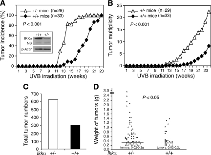
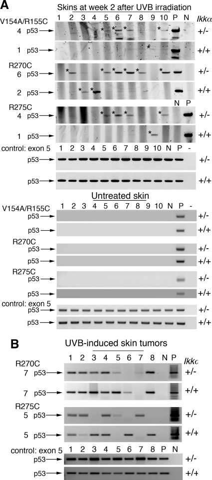
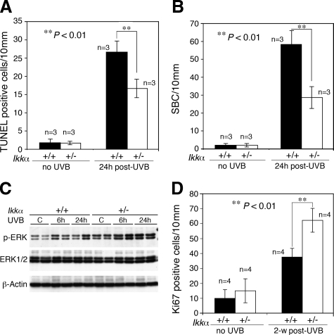
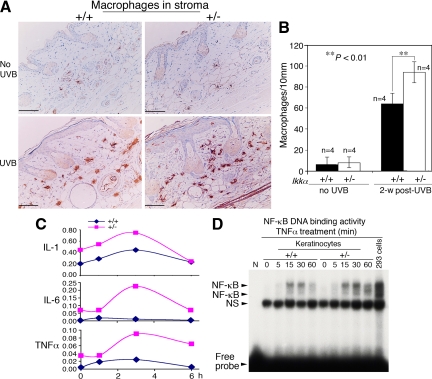
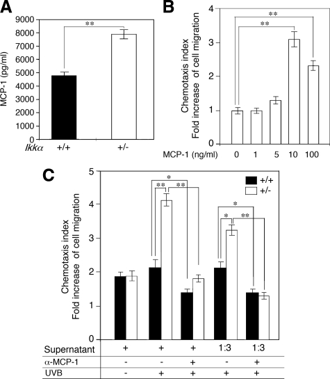
Ultraviolet B light (UVB) is a common cause of human skin cancer. UVB irradiation induces mutations in the tumor suppressor p53 gene as well as chronic inflammation, which are both essential for UVB carcinogenesis. Inhibitor of nuclear factor kappaB kinase-alpha (IKKalpha) plays an important role in maintaining skin homeostasis, and expression of IKKalpha was found to be down-regulated in human and murine skin squamous cell carcinomas. However, the role of IKKalpha in UVB skin carcinogenesis has not been investigated. Thus, here we performed UVB carcinogenesis experiments on Ikkalpha(+/+) and Ikkalpha(+/-) mice. Ikkalpha(+/-) mice were found to develop a twofold greater number of skin tumors than Ikkalpha(+/+) mice after chronic UVB irradiation. In addition, tumor latency was significantly shorter and tumors were bigger in Ikkalpha(+/-) than in Ikkalpha(+/+) mice. At an early stage of carcinogenesis, an increase in UVB-induced p53 mutations as well as macrophage recruitment and mitogenic activity, and a decrease in UVB-induced apoptosis, were detected in Ikkalpha(+/-) compared with those in Ikkalpha(+/+) skin. Also, reduction of IKKalpha levels in keratinocytes up-regulated the expression of monocyte chemoattractant protein-1 (MCP-1/CCL2), TNFalpha, IL-1, and IL-6, and elevated macrophage migration, which might promote macrophage recruitment and inflammation. Therefore, these findings suggest that reduction of IKKalpha expression orchestrates UVB carcinogen, accelerating tumorigenesis.


Figures

Figure 1
Figure 1
Reduction of IKKα expression promotes UVB skin carcinogenesis in mice. A: Skin tumor incidence in Ikkα+/+ (+/+) and Ikkα+/− (+/−) mice (P < 0.001, COX proportional hazards model). IKKα protein levels were detected by Western blotting. β-Actin, loading control. NS indicates nonspecific band. B: Tumor multiplicity in Ikkα+/+ and Ikkα+/− mice (P < 0.001, Poisson regression analysis). C: Tumor numbers in 33 Ikkα+/+ and 29 Ikkα+/− mice at week 28. D: Weight comparison of tumors found in 33 Ikkα+/+ and 29 Ikkα+/− mice (P < 0.05, t test). Lines indicate means; g, Gram (tumor weights).
Figure 2
Figure 2
Reduction of IKKα expression enhances early UVB-induced p53 mutations in mouse skin detected by allele-specific PCR. A: Detection of p53 mutations (V154A/R155C, R270C, R275C) in skins of Ikkα+/+ (+/+) and Ikkα+/− (+/−) mice at week 2 after UVB irradiation and in skins of untreated Ikkα+/+ (+/+) and Ikkα+/− (+/−) mice. V indicates valine; A, alanine; R, arginine; C, cysteine. The numbers on the left represent p53 mutation-positive skins. +/+, Ikkα+/+ skin; +/−, Ikkα+/− skin; *p53 PCR bands with mutations; exon 5, exon 5 of the p53 gene as PCR controls; P, positive control (UVB-induced skin tumors); N, negative control for p53 mutations; −, no DNA in PCR. B: Detection of p53 mutations (R270C; R275C) in skin tumors developed in Ikkα+/+ and Ikkα+/− mice at the end point of the experiment.
Figure 3
Figure 3
Reduction of IKKα expression suppresses UVB-induced apoptosis in mouse skin and promotes cell proliferation. A and B: Paraffin-embedded skin sections obtained from Ikkα+/+ (+/+) and Ikkα+/− (+/−) mice using a TUNEL assay kit and the number of TUNEL-positive cells determined under a microscope. Groups of mice (n = 3) used. The SBCs in the epidermis of hematoxylin and eosin–stained skin sections were counted. 24 hours post-UVB, skin sections prepared from mice at 24 hours after UVB irradiation at 100 mJ/cm2. **P < 0.01, t test. C: Levels of p-ERK and ERK in skin specimens obtained from UVB-irradiated mice detected using Western blotting. β-Actin, protein loading controls; C, untreated sample; 6 hours and 24 hours, samples prepared from mice at 6 hours and 24 hours post-UVB irradiation (100 mJ/cm2). D: Comparison of Ki-67–positive cells immunohistochemically stained in the interfollicle epidermis of mice. Groups of mice (n = 4) used. 2-w post-UVB, skin sections prepared from mice at week 2 after treatment with UVB at 100 mJ/cm2. **P < 0.01, t test.
Figure 4
Figure 4
Reduction of IKKα expression elevates inflammation in the skin. A: Detection of macrophages in Ikkα+/+ (+/+) and Ikkα+/− (+/−) paraffin-embedded skin sections, immunohistochemically stained with a macrophage marker. Brown color, macrophages; blue color, nuclear countingstaining with hemetoxylin; UVB, UVB irradiation for 2 weeks. B: Comparison of numbers of macrophages in the skin stroma of UVB-irradiated Ikkα+/+ (+/+) and Ikkα+/− (+/−) mice shown in A. Groups of mice (n = 4) used. 2-w post-UVB, skin prepared from mice at week 2 after UVB irradiation with 100 mJ/cm2. **P < 0.01, t test. Scale bars = 30 μm. C: Relative levels of TNFα, IL-6, and IL-1 mRNA in UVB-irradiated Ikkα+/+ (+/+) and Ikkα+/− (+/−) primary cultured keratinocytes detected using RT-PCR. Levels of GAPDH mRNA were used to normalize the expression levels of these cytokines. Each point (n = 3) used. 2, 4, 6 hours, primary cultured keratinocytes were collected at 2, 4, and 6 hours after irradiation (100 mJ/cm2). D: NF-κB DNA-binding activity in primary cultured keratinocytes treated with TNFα (10 ng/ml) from 0 to 60 minutes. 293 cells treated with TNFα for 30 minutes were used as a positive control. NS indicates nonspecific band.
Figure 5
Figure 5
Reduction of IKKα in keratinocytes promotes expression of MCP-1 and macrophage migration. A: Comparison of MCP-1 expression in UVB-irradiated primary cultured Ikkα+/+ (+/+) and Ikkα+/− (+/−) keratinocytes at 24 hours after irradiation by using ELISA assay. **P < 0.01, t test. B: Activity of recombinant MCP-1 in inducing macrophage migration. **P < 0.01, t test. C: Comparison of activities of the supernatant of primary cultured Ikkα+/+ (+/+) and Ikkα+/− (+/−) keratinocytes. UVB, collecting cells at 24 hours after UVB treatment at 100 mJ/cm2; α-MCP-1, antibody against MCP-1; 1:3, 1/3 dilution. *P < 0.05, t test; **P < 0.01, t test.

References

    1. Ziegler A, Jonason AS, Leffell DJ, Simon JA, Sharma HW, Kimmelman J, Remington L, Jacks T, Brash DE. Sunburn and p53 in the onset of skin cancer. Nature. 1994;372:773–776. - PubMed
    1. Jonason AS, Kunala S, Price GJ, Restifo RJ, Spinelli HM, Persing JA, Leffell DJ, Tarone RE, Brash DE. Frequent clones of p53-mutated keratinocytes in normal human skin. Proc Natl Acad Sci USA. 1996;93:14025–14029. - PMC - PubMed
    1. Berg RJ, van Kranen HJ, Rebel HG, de Vries A, van Vloten WA, Van Kreijl CF, van der Leun JC, de Gruijl FR. Early p53 alterations in mouse skin carcinogenesis by UVB radiation: immunohistochemical detection of mutant p53 protein in clusters of preneoplastic epidermal cells. Proc Natl Acad Sci USA. 1996;93:274–278. - PMC - PubMed
    1. Rebel H, Mosnier LO, Berg RJ, Westerman-de Vries A, van Steeg H, van Kranen HJ, de Gruijl FR. Early p53-positive foci as indicators of tumor risk in ultraviolet-exposed hairless mice: kinetics of induction, effects of DNA repair deficiency, and p53 heterozygosity. Cancer Res. 2001;61:977–983. - PubMed
    1. Ouhtit A, Gorny A, Muller HK, Hill LL, Owen-Schaub L, Ananthaswamy HN. Loss of Fas-ligand expression in mouse keratinocytes during UV carcinogenesis. Am J Pathol. 2000;157:1975–1981. - PMC - PubMed

Publication types