. 2010 May 18;17(2):513-24.
doi: 10.1677/ERC-09-0272. Print 2010 Jun.

Defective nucleolar localization and dominant interfering properties of a parafibromin L95P missense mutant causing the hyperparathyroidism-jaw tumor syndrome

Affiliations

Defective nucleolar localization and dominant interfering properties of a parafibromin L95P missense mutant causing the hyperparathyroidism-jaw tumor syndrome

Leelamma M Panicker et al. Endocr Relat Cancer. .

Abstract

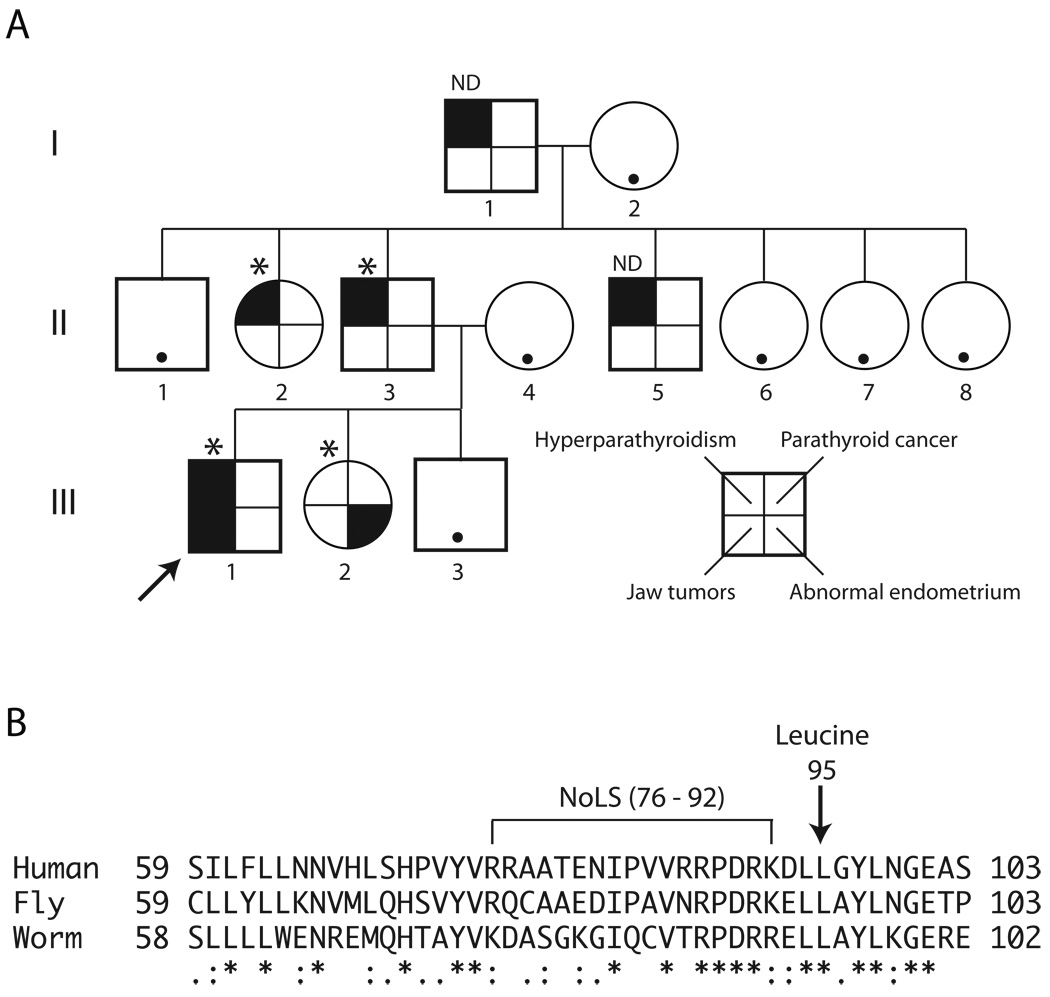
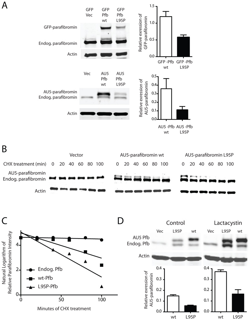
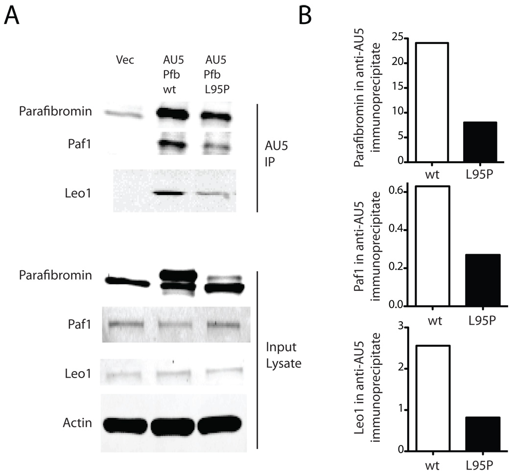
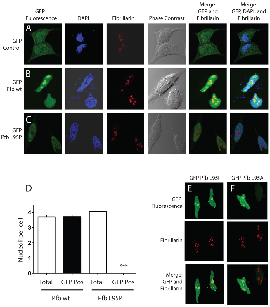
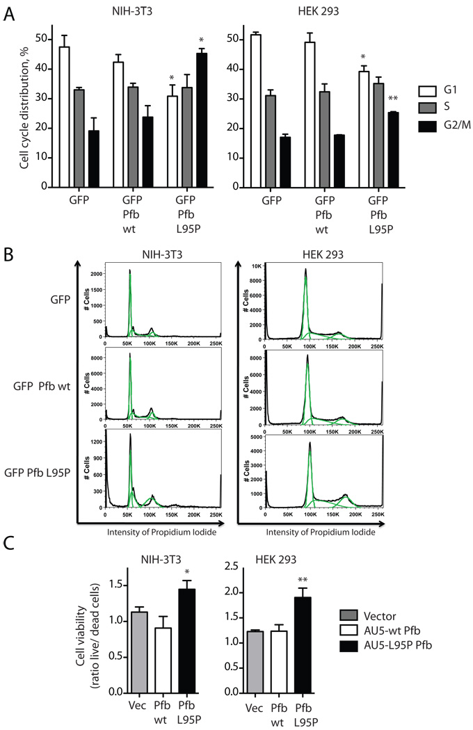
The hyperparathyroidism-jaw tumor syndrome (HPT-JT) is a familial cancer syndrome that can result from germline inactivation of HRPT2/CDC73, a putative tumor suppressor gene that encodes parafibromin, a component of the transcriptional regulatory PAF1 complex with homology to the yeast protein Cdc73p. The vast majority of HRPT2/CDC73 germline mutations identified have been truncation or frameshift mutations, and loss of function due to missense mutation is rare. We report here a kindred with HPT-JT due to a germline L95P missense mutation in parafibromin. The mutant parafibromin was studied in vitro to understand the basis of its presumed loss-of-function. When transfected in cultured cells, the L95P mutant was expressed to a lower level than wild-type (wt) parafibromin, a difference that was not overcome by inhibition of the proteasomal degradation pathway. The L95P mutant parafibromin retained the ability to assemble with endogenous PAF1 complex components as evidenced by co-immunoprecipitation. Analysis of subcellular localization showed that the L95P mutant was markedly deficient in nucleolar localization compared to the wt, an impairment likely resulting from disruption of a putative nucleolar localization signal immediately upstream of the L95P mutation. Transfection of the L95P parafibromin mutant, but not the wt, enhanced cell cycle progression and increased cell survival in NIH-3T3 and HEK 293 cells, resulting apparently from dominant interference with endogenous parafibromin action. The simultaneous loss of nucleolar localization and acquisition of a growth stimulatory phenotype with the L95P mutation raise the possibility that parafibromin must interact with targets in the nucleolus to fully execute its tumor suppressor functions.


Conflict of interest statement

DECLARATION OF INTEREST

None of the authors has a conflict of interest that could be perceived as prejudicing the impartiality of the research reported herein.

Figures

Figure 1
Figure 1. Kindred with the hyperparathyroidism-jaw tumor syndrome associated with germline HRPT2/CDC73 L95P missense mutation
A. Pedigree of study kindred with the hyperparathyroidism-jaw tumor syndrome including three generations of affecteds. Square symbols indicate males, and round symbols indicate females, with legend to phenotypes shown in lower right. The arrow indicates the proband (III-1). The asterisk above a symbol indicates positivity for germline HRPT2/CDC73 L95P missense mutation testing, and ND above a symbol indicates gene testing not yet performed. A black dot within an otherwise blank symbol indicates that the serum calcium was normal at the latest screening in an individual never known to have hypercalcemia. B. Sequence alignment of human parafibromin protein residues 59–103 with the corresponding regions of predicted homologs from Drosophila melanogaster (Hyrax, CG11990) and the nematode Brugia malayi (NCBI Access. No. XP_001895980). The position of the conserved leucine at residue 95 in human parafibromin is indicated (arrow), as is the position of a nucleolar localization signal (NoLS) encompassing residues 76 –92 previously described (Hahn and Marsh 2007). Multiple sequence alignment was generated with ClustalW2 software (v. 2.0.12) available at the European Bioinformatics Institute website (http://www.ebi.ac.uk/Tools/clustalw2/), using the default parameters (Larkin, et al. 2007). “*”, residues in the column are identical in all sequences in the alignment, “:”, conserved substitutions observed in the alignment; “.”, semiconserved substitutions observed in the alignment.
Figure 2
Figure 2. Diminished expression level of the L95P parafibromin mutant not due to accelerated proteasomal degradation
A (Left) Immunoblots of GFP control or GFP fusions with wild-type (wt) or L95P mutant parafibromin (Pfb) (upper panels) and vector control (Vec), or AU5 epitope-tagged wild-type or L95P mutant parafibromin (lower panels) and endogenous parafibromin in cultured Hela cells after 24 hours of transfection. (Right) Histograms quantifying the relative expression level of wild type and L95P mutant parafibromin constructs compared to β-actin in the same sample. B. Time course of protein expression of endogenous parafibromin, AU5-parafibromin and AU5 L95P-parafibromin following cycloheximide treatment to block de novo protein synthesis. Cultured HeLa cells were transfected with the indicated constructs, then 6 hours following transfection cycloheximide was added to the culture medium and cells were collected at 0, 20, 40, 60, 80 and 100 min. Cell lysates were analyzed by immunoblotting at the indicated time points using anti-parafibromin antibody (upper) or anti-actin (lower) antibodies. The expression of β-actin is shown as a loading control. C. Linear regression analysis of the relative expression of transfected and endogenous parafibromin from (B) as described in Experimental Procedures. D. Immunoblots (above) and histograms (below) showing the expression, relative to β-actin, of wild type and L95P mutant AU5-tagged parafibromin without and with treatment with the proteasome inhibitor lactacystin for two hours.
Figure 3
Figure 3. The L95P parafibromin mutant retains interaction with PAF1 complex components
A. 24 hours after transfection with vector only (Vec), or AU5 epitope-tagged wild-type (wt) or L95P mutant parafibromin (Pfb) HeLa cells were immunoprecipitated using AU5 monoclonal antibody and both the starting cell lysate (lower) and washed immunoprecipitates (upper) were analyzed by immunoblotting using anti-parafibromin, anti-Paf1 and anti-Leo1 antibodies as indicated. B. Histograms comparing the amount of immunoreactive parafibromin, Paf1 and Leo1 in the anti-AU5 immunoprecipitates.
Figure 4
Figure 4. The L95P parafibromin mutation blocks nucleolar but not nuclear localization
Hela cells were cultured in chamber slides and transfected with GFP vector control (A) or else GFP fusions with wild-type (Pfb wt) (B) or L95P mutant parafibromin (Pfb L95P) (C). Cells were then treated with the DAPI nuclear stain, immunostained using anti-fibrillarin antibody as a nucleolar marker, and analyzed by confocal laser fluorescence microscopy. Phase contrast and overlay (merge) of GFP fluorescent signal with the nucleolar marker without or with the nuclear stain are also shown. D. Quantitation of nucleoli per Hela cell transfected with GFP fusions with wild-type (Pfb wt) or L95 mutant parafibromin (Pfb L95P): total nucleoli counted if red on fibrillarin-only stained images; GFP-positive nucleoli counted if yellow on merging of GFP fluorescence and fibrillarin stained images. Means shown from pooling of 2 or 3 experiments per transfection, with 19 to 24 cells (nuclei) analyzed per experiment. ***, two-tailed P value <0.0001 compared to total, employing Student’s unpaired t test. E., F. Confocal laser fluorescence microscopic images of Hela cells transfected with GFP fusions with L95I mutant (Pfb L95I) (E) or L95A mutant parafibromin (Pfb L95A) (F), and analyzed as indicated, in A–C.
Figure 5
Figure 5. The L95P parafibromin mutant enhances cell cycle progression and promotes cell survival in vitro
A. Cell cycle analysis. NIH-3T3 cells and HEK 293 cells were transfected with GFP vector cDNA control or else cDNAs encoding fusion proteins of GFP with wild-type (wt) or L95 mutant parafibromin (Pfb). After 48 hours of transfection the cells were subjected to flowcytometric analysis in GFP expressing cells. Histograms show the percentage of cells in the indicated phases of the cell cycle. Values shown represent mean and standard errors of pooled results from of (HEK 293) or 3 (NIH-3T3) independent experiments; **, two-tailed P value <0.02; * <0.05 compared to GFP control, employing Student’s unpaired t test. B. Flowcytometric analysis plotting cell number versus intensity of propidium iodide signal for cells transfected under the indicated conditions, from a representative experiment described in A. C. Cell viability analysis. Cultured NIH-3T3 cells and HEK 293 cells were transfected with vector only (Vec), or AU5 epitope-tagged wild-type (wt) or L95P mutant parafibromin (Pfb) and after 48 hrs were analyzed for cell viability as described in Experimental Procedures. Ordinate values show the ratio of live to dead cells, representing the pooled data of six independent repeats. *, two-tailed P value <0.05; ** <0.01 compared to vector and wild-type controls, employing Fisher’s exact test.

Similar articles

Cited by

References

    1. Bradley KJ, Cavaco BM, Bowl MR, Harding B, Cranston T, Fratter C, Besser GM, Conceicao Pereira M, Davie MW, Dudley N, et al. Parafibromin mutations in hereditary hyperparathyroidism syndromes and parathyroid tumours. Clin Endocrinol (Oxf) 2006;64:299–306. - PubMed
    1. Bradley KJ, Hobbs MR, Buley ID, Carpten JD, Cavaco BM, Fares JE, Laidler P, Manek S, Robbins CM, Salti IS, et al. Uterine tumours are a phenotypic manifestation of the hyperparathyroidism-jaw tumour syndrome. J Intern Med. 2005;257:18–26. - PubMed
    1. Carpten JD, Robbins CM, Villablanca A, Forsberg L, Presciuttini S, Bailey-Wilson J, Simonds WF, Gillanders EM, Kennedy AM, Chen JD, et al. HRPT2, encoding parafibromin, is mutated in hyperparathyroidism-jaw tumor syndrome. Nat Genet. 2002;32:676–680. - PubMed
    1. Cetani F, Pardi E, Borsari S, Viacava P, Dipollina G, Cianferotti L, Ambrogini E, Gazzerro E, Colussi G, Berti P, et al. Genetic analyses of the HRPT2 gene in primary hyperparathyroidism: germline and somatic mutations in familial and sporadic parathyroid tumors. J Clin. Endocrinol. Metab. 2004;89:5583–5591. - PubMed
    1. Chang M, French-Cornay D, Fan HY, Klein H, Denis CL, Jaehning JA. A complex containing RNA polymerase II, Paf1p, Cdc73p, Hpr1p, and Ccr4p plays a role in protein kinase C signaling. Mol Cell Biol. 1999;19:1056–1067. - PMC - PubMed

Publication types

MeSH terms