. 2010 May;24(5):898-913.
doi: 10.1210/me.2009-0310. Epub 2010 Mar 19.

The rules of DNA recognition by the androgen receptor

Affiliations

The rules of DNA recognition by the androgen receptor

Sarah Denayer et al. Mol Endocrinol. 2010 May.

Abstract

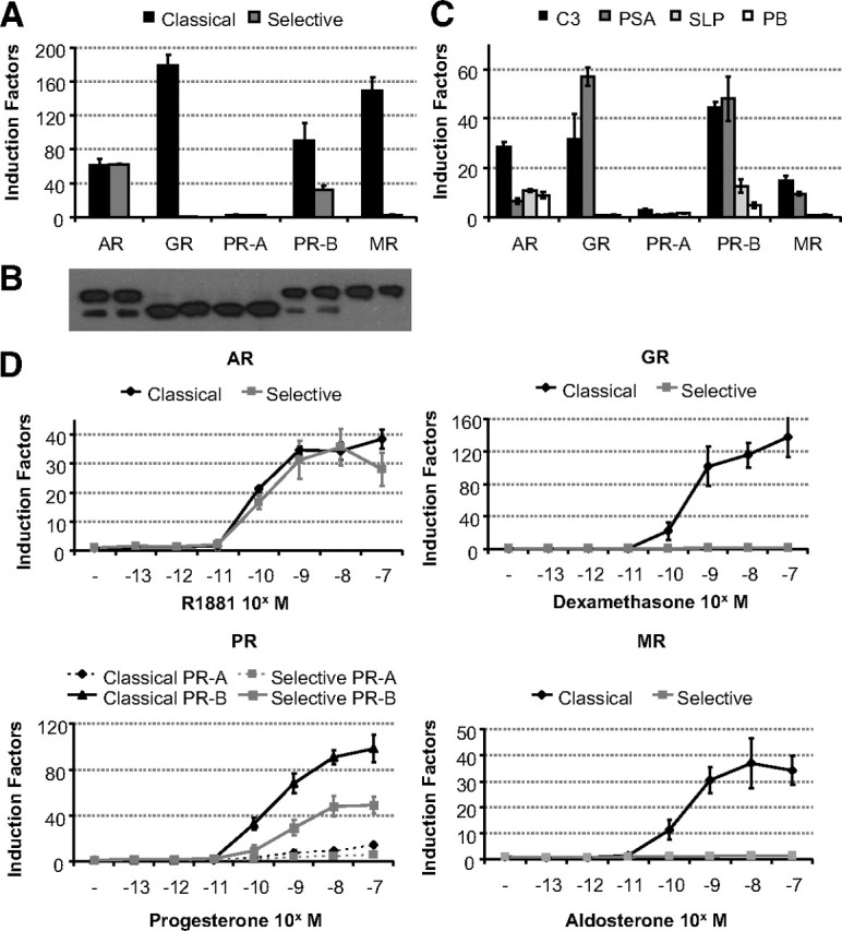
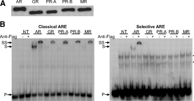
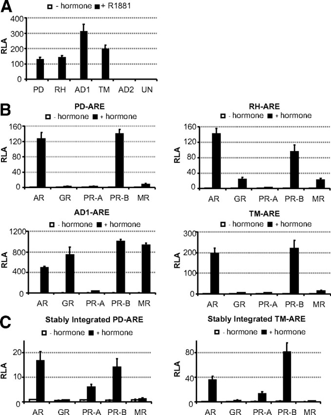
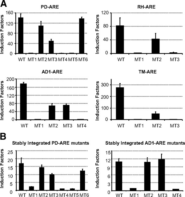
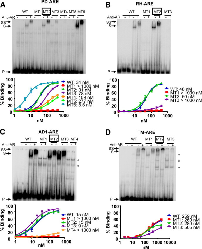
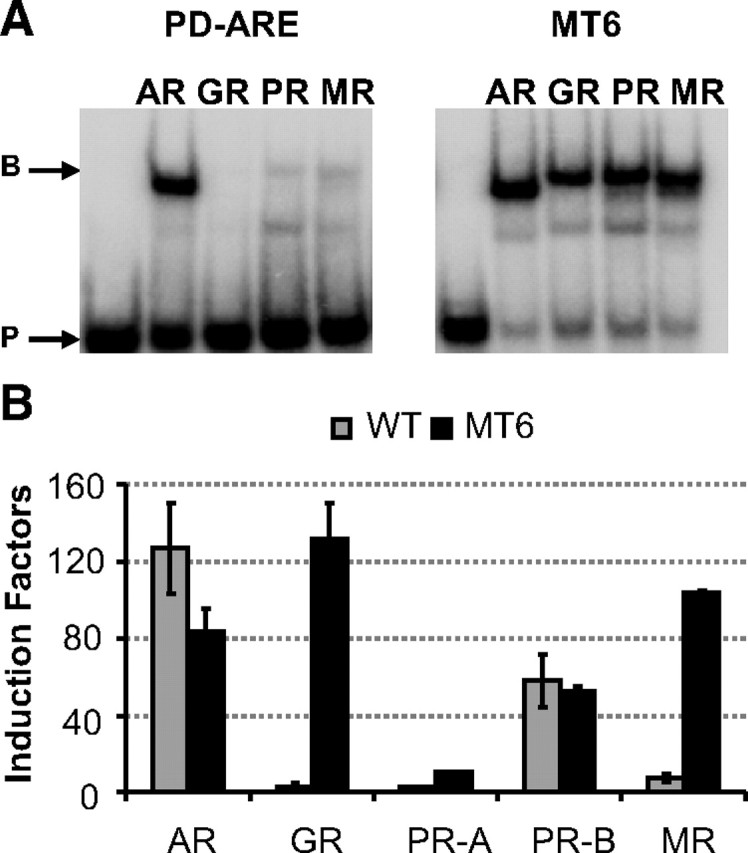
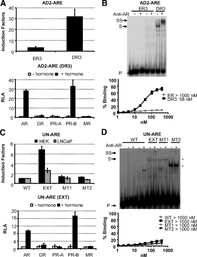
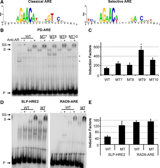
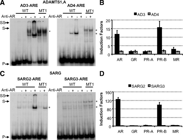
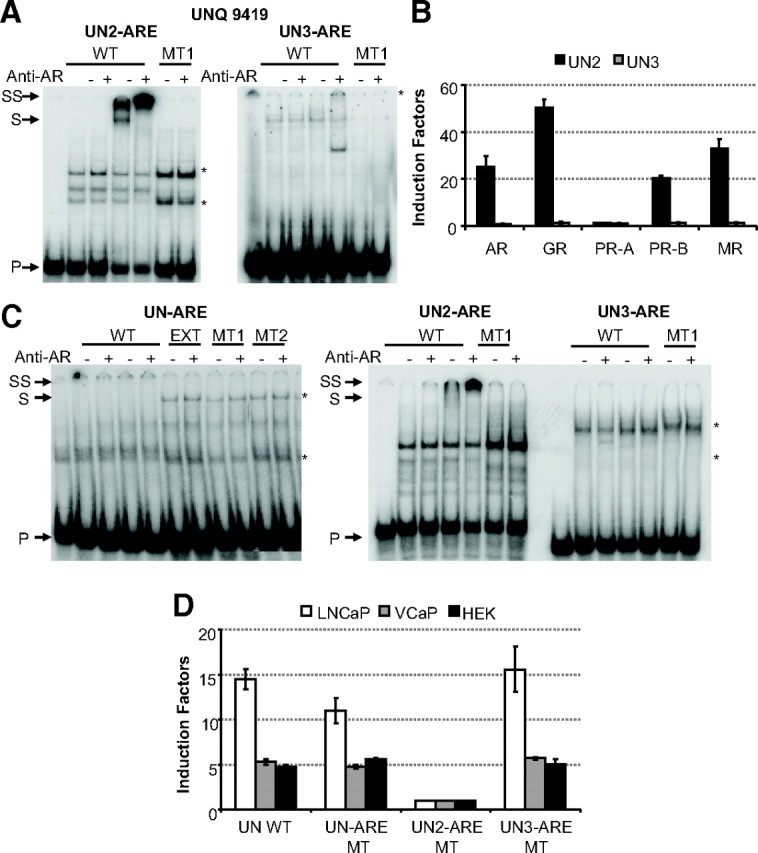
The androgen receptor (AR) and glucocorticoid, progestagen, and mineralocorticoid receptors all recognize classical DNA response elements that are organized as inverted repeats of 5'-AGAACA-3'-like motifs with a three-nucleotide spacer. Next to such elements, the AR also recognizes a second type of androgen response element (ARE), the so-called selective AREs, which resemble more the direct repeats of the same hexamer. In this work, we show that not only the AR but also the progestagen receptor can recognize the selective AREs, whereas neither glucocorticoid nor mineralocorticoid receptor can. Recently, genomic AR-binding fragments have been postulated to contain AR-binding sites that diverge considerably from the classical ARE consensus. Extensive mutational analyses of these candidate motifs, however, reinstalls the values of the consensus sequence for the AREs as mentioned above, the importance of their dimeric nature and the presence of exactly three-nucleotide spacing. We developed a position-specific probability matrix that was used to predict with higher accuracy new AREs in different AR-binding regions. So far, all AR-binding genomic fragments that were analyzed contain AREs defined as receptor-dimer binding motifs with the ability to confer responsiveness to a reporter gene.


Figures

Fig. 1.
Fig. 1.
Comparison of receptor selectivity of classical and selective enhancers and promoters. A, HEK293 cells (104 per 96 wells) were transfected with 100 ng of a classical or a selective ARE-based reporter and 10 ng of a receptor expression vector (see Table 4 for the ARE sequences). Cells were stimulated for 24 h with R1881, dexamethasone, progesterone, or aldosterone (all at 10−8 m). Results are presented as induction factors. Error bars are the averages’ sem of at least three independent experiments performed in triplicate. B, For immunoblotting, transfected HEK293 cells were treated for 1 h without or with 10 nm of the relevant hormone. The expressed proteins were detected using an anti-Flag antibody. C, HEK293 cells were transfected as in A with 100 ng of a reporter gene driven by a single copy of the classical C3(1 )-enhancer and PSA-promoter or the selective slpARU-TK-TATA and probasin promoter. Cells were stimulated for 24 h with 10 nm R1881, dexamethasone, progesterone, or aldosterone. Results are presented as in A. D, HEK293 cell lines containing a stable integrated classical or selective ARE were made using the Flp-In T-REx system (Invitrogen). The indicated receptor expression vector (100 ng) was transiently transfected. Cells were stimulated for 24 h with different concentrations of R1881, dexamethasone, progesterone, or aldosterone. Results are presented as in A.
Fig. 2.
Fig. 2.
EMSA of nonselective and selective AREs by full-size receptors. A, COS-7 cells were transfected with 8 μg receptor expression plasmid. Cells were treated for 1 h with 10 nm R1881, dexamethasone, progesterone, or aldosterone. Nuclear extracts were made as described in Materials and Methods. The expressed proteins were blotted and detected using the M2 anti-Flag antibody. B, 32P-labeled classical (SLP-MUT) or selective (SLP-HRE) probes were incubated with similar amounts of nuclear extract from nontransfected (NT) COS-7 cells or nuclear extracts containing the AR, GR, PR-A, PR-B, or MR. No protein was added in the first lane as a negative control. Anti-Flag antibody was added as indicated at the top. Arrows indicate the positions of the unbound (P), shifted (S), and supershifted (SS) probe. Asterisks indicate nonspecific complexes.
Fig. 3.
Fig. 3.
Functional analysis of candidate AREs. A, HEK293 cells were transfected with 100 ng reporter constructs of the indicated AREs and cotransfected with 10 ng AR expression plasmid. Cells were incubated for 24 h in medium without or with hormone (10 nm R1881). Results are presented as relative luciferase activity. Error bars are the averages’ sem of at least three independent experiments performed in triplicate. B, Identified AREs were classified as classical or selective AREs. Transfection assays were performed as in Fig. 1A. C, HEK293 cells stably integrated the PD- or TM-ARE were made to verify the effect on chromatin. Cells were transfected with 100 ng receptor expression plasmid and stimulated after 24 h with R1881, dexamethasone, progesterone, or aldosterone (10−8 m). Results are presented as relative luciferase activity (RLA). Error bars are the averages’ sem of at least three independent experiments performed in triplicate.
Fig. 4.
Fig. 4.
Mutational analysis of the ARE spacer lengths: functional studies. A, HEK293 cells stably expressing the AR were transiently transfected with 100 ng reporter plasmid as described in Materials and Methods. Cells were stimulated for 24 h without or with hormone (10 nm R1881). Bars represent induction factors. Error bars are the averages’ sem of at least three independent experiments performed in triplicate. The sequences of the different DNA elements are given in Table 1. B, HEK293 cell lines containing a stable integrated wild-type (WT) or mutant PD- or AD1-ARE were made using the Flp-In T-REx system (Invitrogen). The AR expression vector (100 ng) was transiently transfected. Cells were treated for 24 h without or with 10 nm. Results are presented as in A.
Fig. 5.
Fig. 5.
Mutational analysis of variable spacer lengths of PD-ARE (A), RH-ARE (B), AD1-ARE (C), and TM-ARE (D) by EMSA with full-length AR and isolated AR-DBD. Probes were incubated with nuclear extracts before electrophoresis as described in Materials and Methods. In the EMSA experiments shown, the first five lanes contain the wild-type (WT) probes; in lanes 2–5, these probes were incubated with COS-7 extracts without (lanes 2 and 3) or with (lanes 4 and 5) AR. The sequences of the different wild-type and mutant probes tested are given in Table 1. Anti-AR antibody was added as indicated at the top. Arrows indicate the positions of the unbound (P), shifted (S), and supershifted (SS) probe. Mutants testing variable spacer lengths are boxed. Asterisks indicate nonspecific complexes. The graphs in the lower panels result from EMSA with isolated AR-DBDs. The amount of radioactivity present in dimer-bound DNA was calculated relative to the total amount of radioactivity in each lane and plotted against the concentration of protein that was used. Fits were calculated to a curve with Hill kinetics, and apparent dissociation constants (Kd values) were calculated.
Fig. 6.
Fig. 6.
Effect of changing the direct repeat nature of the PD-ARE. Panel A, 32P-labeled PD-ARE or MT6 (Table 1) was incubated with 200 ng AR-, GR-, PR-, or MR-DBD (Table 1). In the first lane, no protein was added as a negative control. Arrows indicate the position of the unbound probe (P) and bound probe (B). Panel B, HEK293 cells were transfected with 100 ng reporter plasmids containing the PDE9A wild-type (WT) or MT6 ARE and 10 ng receptor expression plasmid. Cells were incubated for 24 h without or with hormone (10 nm R1881, dexamethasone, progesterone, or aldosterone). Results are represented as induction factors. Error bars are the averages’ sem of at least three independent experiments performed in triplicate.
Fig. 7.
Fig. 7.
Mutational analysis of alternative hexamer orientations in AD2-ARE (A and B) and UN-ARE (C and D). A functional analysis was performed in HEK293 cell stably expressing the AR (A and C, upper panels). The sequences of the everted repeat (ER3) and direct repeat (DR3) for the AD2-ARE are given in Table 1 as well as the wild-type (WT) UN-ARE, the extended UN-ARE and mutants of the latter. Reporter constructs (100 ng) were transiently cotransfected. Cells were treated without or with 10 nm R1881 for 24 h. Results are given as induction factors. The lower panels of A and C test the selectivity of the DR3 (AD2-ARE) and EXT (extended UN-ARE) as described in the legend of Fig. 1A. DNA-binding studies were performed as described in the legend of Fig. 5. Arrows indicated the positions of the unbound (P), shifted (S), and supershifted (SS) probe. Asterisks indicate nonspecific complexes. The lower panels of B and D result from EMSA with AR-DBDs. Apparent dissociation constants (Kd values) were calculated as described in the legend of Fig. 5.
Fig. 8.
Fig. 8.
Nucleotides flanking the high-affinity binding site. A, Sequence logos illustrating sequence conservation for classical and selective AREs were made based on the sequences shown in Table 4 (http://weblogo.berkeley.edu/). Letter heights represent the degree of base conservation at that position. B and D, 32P-labeled probes were incubated with nuclear extracts before electrophoresis. The first five lanes contain the wild-type (WT) PD-, SLP-, or RAD9A-ARE probe; in lanes 2–5, these probes were incubated with COS-7 nuclear extract without (lanes 2 and 3) or with (lanes 4 and 5) AR. The sequences of the different probes and mutants tested are given in Table 2. Anti-AR antibody was added as indicated at the top. Arrows indicate the positions of the unbound (P), shifted (S), and supershifted (SS) probe. C and E, Reporter constructs containing the wild-type or mutant AREs were transfected in HEK293 cells stably expressing the AR as described in the legend of Fig. 4. *, P < 0.001.
Fig. 9.
Fig. 9.
Characterization of predicted AREs in the ADAM1 enhancer (A and B) and SARG enhancer (C and D). A and C show EMSA experiments to investigate binding of the full-length AR to the predicted AREs found in the ADAM1 enhancer (A) and SARG enhancer (C). The first five lanes contain the wild-type (WT) ARE probe; in lanes 2–5, these probes were incubated with COS-7 extract without (lanes 2 and 3) or with (lanes 4 and 5) AR. Lanes 6 and 7 test AR binding to a mutant ARE. The sequences of the wild-type probes are given in Table 5. Anti-AR antibody was added as indicated at the top. Arrows indicate the positions of the unbound (P), shifted (S), and supershifted (SS) probe. Asterisks indicate nonspecific complexes. B and D display a functional analysis of the predicted AREs in which they are classified as classical and selective AREs. HEK293 cells were transfected with 100 ng reporter construct, and 10 ng receptor expression plasmids were cotransfected. Cells were treated for 24 h without or with hormone (10 nm R1881, dexamethasone, progesterone, or aldosterone). Results are given as induction factors. Error bars are the averages’ sem of at least three independent experiments performed in triplicate.
Fig. 10.
Fig. 10.
Determination of the primary ARE in the UNQ 9419 promoter. A, EMSA experiment to investigate binding of the full-length AR to the predicted AREs found in the UNQ promoter. The first five lanes contain the wild-type (WT) ARE probe; in lanes 2–5, these probes were incubated with COS-7 extract without (lanes 2 and 3) or with (lanes 4 and 5) AR. Lanes 6 and 7 test AR binding to a mutant ARE. The sequences of the wild-type probes are given in Table 5. Anti-AR antibody was added as indicated at the top. Arrows indicate the positions of the unbound (P), shifted (S), and supershifted (SS) probe. Asterisks indicate nonspecific complexes. B, Functional analysis of the predicted AREs in which they are classified as classical and selective AREs as described in Fig. 9. C, EMSA experiment to investigate binding of the full-length AR to the three possible AREs found in the UNQ promoter using VCaP nuclear extracts. The first five lanes contain the wild-type ARE probe; in lanes 2–5, these probes were incubated with nonstimulated VCaP extracts (lanes 2 and 3) or with R1881-stimulated VCaP extracts (lanes 4 and 5). In the left panel, lanes 6–11 test AR binding to the extended UN-ARE and mutants of the latter. In the middle panel, lanes 6 and 7 test AR binding to a mutant ARE. The sequences of the wild-type probes are given in Table 1 or 5. Anti-AR antibody was added as indicated at the top. Arrows indicate the positions of the unbound (P), shifted (S), and supershifted (SS) probe. Asterisks indicate nonspecific complexes. D, HEK293, LNCaP, and VCaP cells were transfected with reporter constructs containing the wild-type or mutant UNQ promoter fragment. Cells were treated without or with 10 nm R1881 for 24 h. Results are represented as induction factors. Error bars are the averages’ sem of at least three independent experiments performed in duplicate.

Similar articles

Cited by

References

    1. Evans RM1988. The steroid and thyroid hormone receptor superfamily. Science 240:889–895 - PMC - PubMed
    1. Forman BM, Samuels HH1990. Interactions among a subfamily of nuclear hormone receptors: the regulatory zipper model. Mol Endocrinol 4:1293–1301 - PubMed
    1. Glass CK1994. Differential recognition of target genes by nuclear receptor monomers, dimers, and heterodimers. Endocr Rev 15:391–407 - PubMed
    1. Mangelsdorf DJ, Thummel C, Beato M, Herrlich P, Schütz G, Umesono K, Blumberg B, Kastner P, Mark M, Chambon P, Evans RM1995. The nuclear receptor superfamily: the second decade. Cell 83:835–839 - PMC - PubMed
    1. Claessens F, Alen P, Devos A, Peeters B, Verhoeven G, Rombauts W1996. The androgen-specific probasin response element 2 interacts differentially with androgen and glucocorticoid receptors. J Biol Chem 271:19013–19016 - PubMed

Publication types