. 2010 Apr 1;9(7):1421-33.
doi: 10.4161/cc.9.7.11256. Epub 2010 Apr 1.

Identification of Filamin A as a BRCA1-interacting protein required for efficient DNA repair

Affiliations

Identification of Filamin A as a BRCA1-interacting protein required for efficient DNA repair

Aneliya Velkova et al. Cell Cycle. .

Abstract

The product of the breast and ovarian cancer susceptibility gene BRCA1 has been implicated in several aspects of the DNA damage response but its biochemical function in these processes has remained elusive. In order to probe BRCA1 function we conducted a yeast two-hybrid screening to identify interacting partners to a conserved motif (Motif 6) in the central region of BRCA1. Here we report the identification of the actin-binding protein Filamin A (FLNA) as BRCA1 partner and demonstrate that FLNA is required for efficient regulation of early stages of DNA repair processes. Cells lacking FLNA display a diminished BRCA1 IR-induced focus formation and a delayed kinetics of Rad51 focus formation. In addition, our data also demonstrate that FLNA is required to stabilize the interaction between components of the DNA-PK holoenzyme, DNA-PKcs and Ku86 in a BRCA1-independent fashion. Our data is consistent with a model in which absence of FLNA compromises homologous recombination and non-homologous end joining. Our findings have implications for the response to irradiation induced DNA damage.


Figures

Figure 1
Figure 1
Interaction of Filamin A and BRCA1 in mammalian cells. (A) Left, co-immunoprecipitation of endogenous BRCA1 with FLNA in HeLa and HCT116 cells showing interaction of endogenous BRCA1 and FLNA. Right, reverse reaction showing co-immunoprecipitation of endogenous FLNA with BRCA1. Vertical lines indicate where lanes were digitally removed. Figures are not a composite of lanes from two different blots but from the same blot. (B) Left, diagram of deletion constructs used to map the BRCA1 interaction site to FLNA. RING, RING finger domain; NLS, nuclear localization signals; BRCT, BRCA1 C-terminal domains. Right, co-expression of GST-fragments of BRCA1 and FLAG-FLNA (aa 2477-2647) in 293FT cells. Lower molecular weight band obtained in the empty vector (V) transfection corresponds to GST. (C) Co-immunoprecipitation of BRCA1 fragments (WB GST), endogenous BRCA1 (WB BRCA1) with FLAG-FLNA (aa 2477-2647) under low (left panel) and high (right panel) stringency conditions. Note that endogenous FLNA is also immunoprecipitated by FLAG-FLNA (aa 2477-2647) confirming it is in the native conformation (WB FLNA). Strong reactivity shown for GST BF1 is due to recognition of the GST-BRCA1 fragment that contains the epitope for the antibody. (D) GST-pull down experiments show that GST-BRCA1 fragments 1, 3 and 4 can precipitate endogenous FLNA (WB FLNA).
Figure 2
Figure 2
Fine mapping of the interaction regions in BRCA1. (A) Upper, diagram of deletion constructs used to map the interaction site to FLNA. NES, nuclear export sequence (black boxes). Lower, FLAG-FLNA (aa 2477-2647) interacts strongly with GST-BRCA1 fragments BF1 (aa 1-324), BF1D (aa 141-240) and BF1F (aa 1-302). (B) First, diagram of deletion constructs of fragment aa 141-240 used to map the interaction site to FLNA. The location of the missense variant Y179C is indicated. Second, FLAG-FLNA (aa 2477-2647) co-immunoprecipitates with GST-BRCA1 fragments BF1D1 (aa 160-190) and BF1D3 (aa 160-210). V, GST; D1, GST-BRCA1 fragment BF1D1 (aa 160-190); D2, GST-BRCA1 fragment BF1D2 (aa 190-210); D3, GST-BRCA1 fragments BF1D3 (aa 160-210). Third, control for expression levels. Fourth, GST-BRCA1 fragments BF1D1 (aa 160-190) and BF1D3 (aa 160-210) can pull down endogenous FLNA. (C) Introduction of BRCA1 Y179C mutation significantly reduces BRCA1 interaction to FLAG-FLNA aa 2477-2647 (Left) and to endogenous FLNA (Middle). W, wild type GST-BRCA1 fragment BF1D (aa 141-240); Y, GST-BRCA1 fragment BF1D with Y179C mutation. Right, Introduction of BRCA1 Y179C mutation into a full length BRCA1 context significantly reduces interaction to endogenous FLNA. W, wild type full length BRCA1; Y, full length BRCA1 carrying a Y179C mutation.
Figure 3
Figure 3
FLNA-null cells are deficient in repair but show no impairment in activating the DNA damage response. (A) FLNA+ (A7) and FLNA- (M2) cells were irradiated with 8 Gy or mock-treated (U) and harvested at the indicated time points. While FLNA+ cells repair most of the DSBs (as measured by γ-H2AX) by 8 h, FLNA- cells show significant unrepaired DSBs even after 32 h post-IR (Top). Bottom, shows total levels of H2AX as a loading control. (B) FLNA+ (A7) and FLNA- (M2) cells were irradiated with 8 Gy or mock-treated (NO IR) and harvested at the indicated time points and comet assays were performed under neutral conditions. A two-tailed Student's t test was performed and p values are shown for statistically significant differences. (C) Top two, ATM activation as measured by phosphorylation of S1981 is not compromised in FLNA- cells. Blot for total ATM is used as a loading control. Note significantly higher levels of pS1981-ATM in FLNA- cells. Middle three, no significant difference was observed in levels of DNA-PKcs S2056 or S2609 phosphorylation but recruitment of DNA-PKcs to chromatin is defective in FLNA- cells. Bottom two, ATR presence in chromatin (CHR) is shown. Blot for ATR levels in whole cell lysates is used a loading control. (D) CHK2, CHK1 and NBS1 activation as measured by pT68-CHK2, pS317-CHK1 and pS343-NBS1, respectively is not compromised in FLNA- cells. Note consistently higher levels of pT68-CHK2, pS317-CHK1 and pS343-NBS1 in FLNA- cells. β-actin is used a loading control.
Figure 4
Figure 4
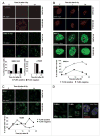
Recruitment of DNA damage response factors to IR-induced foci. (A) Early markers of DNA damage γ-H2AX (red) and phosphoserine 343 NBS1 (green) form foci irrespective of FLNA status. Note maintenance of foci after 24 h only in FLNA- cells. Lower, show quantification of foci-positive cells (≥20 foci). (B) Recruitment of DNA damage response mediator proteins 53BP1 (red, top) and MDC1 (green, middle) was also comparable at early time points. BRCA1 foci formation was compromised in FLNA-deficient cells (lower). (C) Recruitment of repair factor p34 RPA (green, top) did not show any difference between the cell lines at the early time points. FLNA-deficient cells displayed a delayed kinetics of Rad51 foci formation (bottom). In (A–C) representative results from two independent experiments are shown. In each experiment one slide was scored per time point with at least 50 (A) or 100 (B and C) cells scored per slide. Mock-treated cells are indicated by (U). (D) FLNA-deficient cells present with large chromatin-bound RPA foci at 24 h after IR. Higher magnification of FLNA+ and FLNA- cells after 24 h post-IR. Left, shows FLNA+ cells stained for RPA (green). Middle, shows FLNA- cells stained for RPA (green). Note that nuclear foci are significantly larger than in FLNA+ cells. Right, shows a blow up of the inset (white square in middle) with staining for DAPI (blue), RPA (green) and γ-H2AX (red).
Figure 5
Figure 5
Expression of BRCA1-interacting fragment of FLNA or FLNA-interaction fragment of BRCA1 phenocopies loss of FLNA. (A) FLNA- cells were transfected with an empty FLAG vector or a FLAG FLNA-Bf constructs. Cells were mock-treated (U) or treated with 8 Gy IR and cells were collected at different time points. Expression of FLAG FLNA-Bf was unable to reverse the recovery defect. (B) FLNA+ cells were transfected with empty FLAG vector or a FLAG FLNA-Bf constructs. Cells were mock-treated (U) or treated with 8 Gy IR and cells were collected at different time points. Cells expressing of FLAG FLNA-Bf displayed a phenotype similar to FLNA- cells. (C) HCT166 cells stably expressing FLAG FLNA-Bf displayed a phenotype similar to FLNA- cells. (D) FLNA+ cells were transfected with a GST BRCA1-Ff or a GST BRCA1-Ff Y179C. Cells were mock-treated (U) or treated with 8 Gy IR and cells were collected at different time points. Only cells expressing of GST BRCA1-Ff but not GST BRCA1-Ff Y179C displayed a phenotype similar to FLNA- cells, confirming that the effect is specific.
Figure 6
Figure 6
FLNA interacts in vivo with DNA-PKcs and mediates its interaction to Ku86. (A) Left, 293FT cells were transfected with FLAG-FLNA aa 2477-2647 (F) or empty FLAG vector (V) and mock-treated or irradiated with 20 Gy. Cells were collected after 1 h and immunoprecipitated using α-FLAG antibody. FLAG-FLNA aa 2477-2647 co-immunoprecipitates DNA-PKcs in the presence and absence of IR. Right, control for expression and the efficiency of the immunoprecipitation. (B) The interaction between DNA-PKcs and Ku86 is compromised in cells lacking FLNA. Left, shows that levels of DNA-PKcs and Ku86 are similar in both cell lines and in the presence and absence of irradiation. Right, shows that Ku86 and DNA-PKcs interact in FLNA+ cells in the absence of damage and complex formation is significantly increased in the presence of irradiation. Complex formation in the presence and absence of IR is severely compromised in FLNA- cells. (C) Loading of Ku86 onto chromatin after DNA damage is increased in FLNA- cells. Histone H2AX levels are used as a loading control.

Comment in

References

    1. Jackson SP, Bartek J. The DNA-damage response in human biology and disease. Nature. 2009;461:1071–8. - PMC - PubMed
    1. Harper JW, Elledge SJ. The DNA damage response: ten years after. Mol Cell. 2007;28:739–45. - PubMed
    1. Falck J, Coates J, Jackson SP. Conserved modes of recruitment of ATM, ATR and DNA-PKcs to sites of DNA damage. Nature. 2005;434:605–11. - PubMed
    1. Raderschall E, Golub EI, Haaf T. Nuclear foci of mammalian recombination proteins are located at single-stranded DNA regions formed after DNA damage. Proc Natl Acad Sci USA. 1999;96:1921–6. - PMC - PubMed
    1. San Filippo J, Sung P, Klein H. Mechanism of eukaryotic homologous recombination. Annu Rev Biochem. 2008;77:229–57. - PubMed

Publication types

MeSH terms