Comparative Study
. 2010 Apr;13(4):513-20.
doi: 10.1038/nn.2518. Epub 2010 Mar 21.

Monitoring neural activity with bioluminescence during natural behavior

Affiliations
Comparative Study

Monitoring neural activity with bioluminescence during natural behavior

Eva A Naumann et al. Nat Neurosci. 2010 Apr.

Abstract

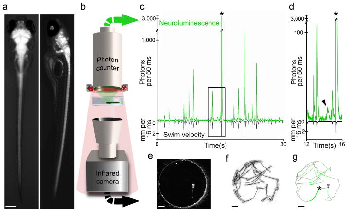
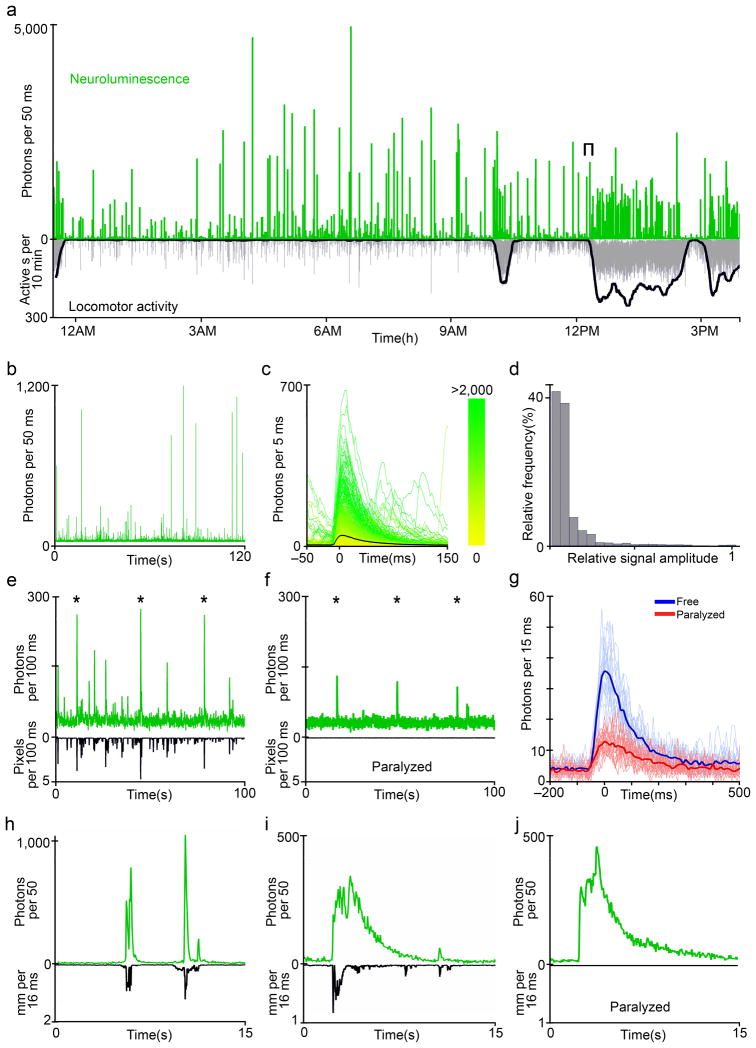
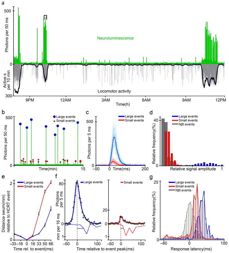
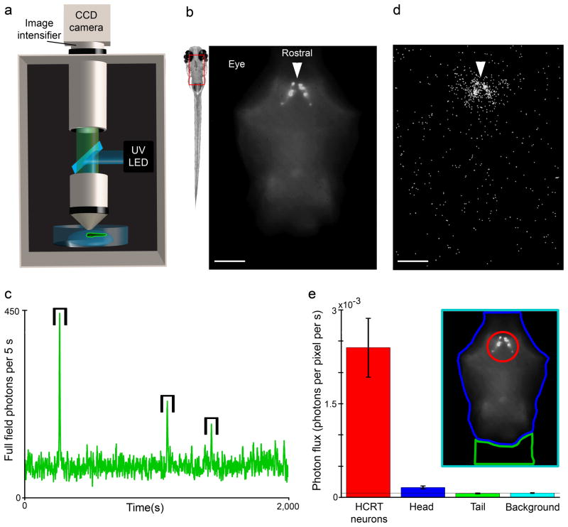
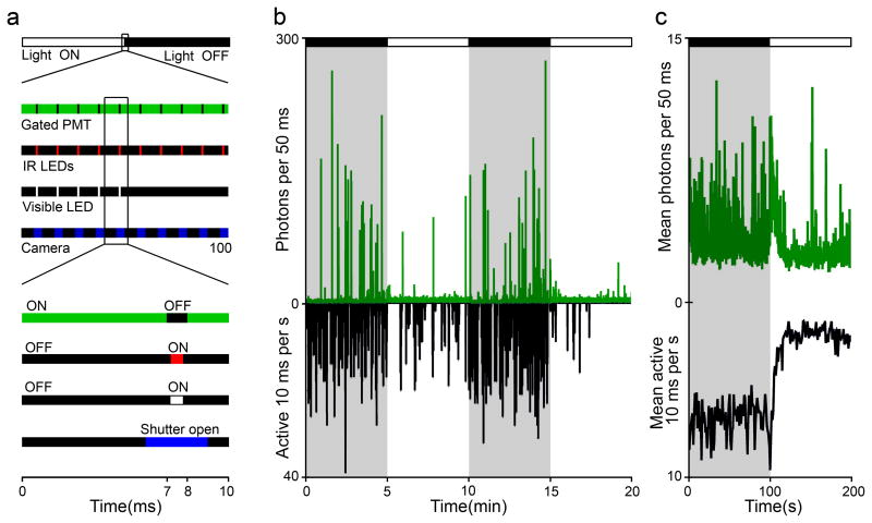
Existing techniques for monitoring neural activity in awake, freely behaving vertebrates are invasive and difficult to target to genetically identified neurons. We used bioluminescence to non-invasively monitor the activity of genetically specified neurons in freely behaving zebrafish. Transgenic fish with the Ca(2+)-sensitive photoprotein green fluorescent protein (GFP)-Aequorin in most neurons generated large and fast bioluminescent signals that were related to neural activity, neuroluminescence, which could be recorded continuously for many days. To test the limits of this technique, we specifically targeted GFP-Aequorin to the hypocretin-positive neurons of the hypothalamus. We found that neuroluminescence generated by this group of approximately 20 neurons was associated with periods of increased locomotor activity and identified two classes of neural activity corresponding to distinct swim latencies. Our neuroluminescence assay can report, with high temporal resolution and sensitivity, the activity of small subsets of neurons during unrestrained behavior.


Figures

Figure 1
Figure 1. Monitoring the neural activity of freely behaving zebrafish
a Dorsal (left) and lateral (right) fluorescence/bright-field micrographs of a 7 dpf Nβt:Ga transgenic zebrafish larva (scale bar: 0.20 mm). b Neuroluminescence setup: A large-area (25 mm diameter) photon-counting PMT is situated above a transparent behavior chamber (12.5 mm diameter). The highly-sensitive light detector is protected by an infrared (IR)-blocking filter such that a ring of 880 nm light-emitting diodes can be used to illuminate the behavioral chamber. Fish are imaged with an IR-sensitive CCD camera positioned below the chamber. The large spectral separation between GA bioluminescence and the IR illumination allows the simultaneous recording of neuroluminescence signals and the behavior of freely swimming zebrafish larvae. c Exemplary neuroluminescence recording of a 7 dpf Nβt:Ga transgenic zebrafish larva previously exposed to coelenterazine. Photon emission and behavior (swim speed in millimeter moved per frame period (mm/16.67 ms)) are shown for a 30 second recording. A mechanical stimulus was delivered at 15 s (*), inducing a fast startle response and a large increase in neuroluminescence. d An expanded view of the boxed region indicated in c highlights a neuroluminescence event not associated with locomotion (arrowhead). e Raw image acquired by the IR CCD camera during neuroluminescence recording (scale bar: 1.5 mm). f Superposition (inverted grayscale) of all frames acquired during the 30 second recording period shown in c, the entire fish trajectory is shown. g The fish trajectory shown in f is overlaid with a colored line for which the neuroluminescence amplitude at each segment is coded as the line-width, (*) indicates the time of the mechanical stimulus.
Figure 2
Figure 2. Neuroluminescence and behavior of NβT GA zebrafish
a Neuroluminescence signals and behavior can be monitored continuously for several days; a 16 hour excerpt of the recording from a 6 dpf Nβt:Ga transgenic zebrafish, following 24 hours of exposure to coelenterazine, is shown. Despite the constant dark conditions of the assay, an increase in locomotor activity, measured as the number of active seconds in a ten minute sliding window (bold line), and a corresponding increase in neuroluminescent events occurs soon after the previously experienced light-on time (9 AM) of the zebrafish light-dark rearing cycle. This is expected from a circadian modulation of spontaneous swimming. b Expanding the bracketed region indicated in a reveals the range of neuroluminescence signal amplitudes that occur during spontaneous behavior. c Upon aligning all the signals detected during the 16 hour recording to each signals onset time and color coding each event by the number of photons arriving in a 50 ms window (0 to >2,000, see color bar), we find that neuroluminescence events consist of a fast rise and slower decay in light emission with a large range of peak amplitudes. d The histogram of signal amplitudes observed from NβT GA zebrafish (n = 6 fish, 3,125 events), normalized to the maximum signal detected from each individual, demonstrates the frequent occurrence of small amplitude events and a long tail of the distribution populated by increasingly large and rare events. e A mechanical stimulus was delivered to a group of freely swimming zebrafish (n = 6) by tapping the recording chamber (stimulus times indicated by the star symbol (*)). The stimulus resulted in neuroluminescence signals coincident with the evoked startle responses, surrounded by intermittent and variable spontaneous signals. f The same fish shown in e were paralyzed with α-Bungarotoxin and received the same mechanical stimulus (*). Paralysis permitted isolating the sensory component of the neuroluminescence event from the full escape response behavior elicited in freely-swimming animals. g The aligned stimulus-driven events in each condition are compared, revealing an attenuated but clearly detectable sensory signal in paralyzed zebrafish. h PTZ induced epileptic behavior, characterized by uncoordinated rapid swimming, is associated with large, fast bursts of neuroluminescence consistent with the strong neural activation expected during seizure episodes (t0 = 1 min after initial PTZ exposure). i Following extended exposure to PTZ (t0 > 17 min), long, slow neuroluminescence events are observed independent of swimming. j Paralyzed zebrafish exposed to PTZ also exhibit long, slow neuroluminescence events, suggesting that motor activity may modulate the amplitude and timescale of PTZ induced epileptic episodes.
Figure 3
Figure 3. Targeted Ga expression in Hypocretin neurons
a Expression of Ga in the ~20 Hypocretin (HCRT) neurons of a transgenic 4 dpf zebrafish larva are imaged with a wide-field fluorescence microscope, demonstrating their position within the posterior diencephalon (scale bar: 100 μm). b Ga-expressing HCRT neurons shown in a maximum intensity projection of image sections acquired with a two-photon microscope (imaged region indicated by red rectangle in a); note the long, dorsal-caudal projecting axons with an expansive arborization near the zebrafish otic vesicle (scale bar: 50 μm).
Figure 4
Figure 4. Activity in Hypocretin neurons during natural behavior
a A freely behaving 4 dpf zebrafish larva exhibits periods of increased spontaneous locomotor activity. The longest active period occurs soon after the light-on time (9 AM) of the normal rearing light cycle. Neuroluminescence events primarily occur during these periods of heightened activity. b Expanding the bracketed region indicated in a reveals that these neural signals fall into two distinct amplitude classes. Manually determined thresholds (200 photons/50 ms in b) were used to classify individual events into a large and small amplitude group. c The amplitude classified signals from the entire recording of the larva shown in a are aligned and the thick lines indicate the average signal time course within each class. d Histogram of the amplitudes for all HCRT neuroluminescence events (n = 1,064, 8 fish), normalized to the maximum response within each fish, are compared to the response amplitude of NβT:GA fish (NβT) shown in Figure 2d. Signals classified as large and small are colored accordingly and are clearly distinct. e The mean distance swum, aligned to the position of the fish at the time of a HCRT signal (0 ms), is plotted for the frames immediately before and after HCRT signals of each amplitude class (error-bars represent s.e.m.). Notably, fish swim sooner and further following small HCRT events than following large HCRT events. f A double exponential fit of neuroluminescence signals was used to identify the peak of the event. Example fits (solid curves) are shown for events (open circles) from the two amplitude classes along with the corresponding swim-velocities. Latency was measured as the time from the peak of the response to time at which the zebrafish achieved a threshold swim velocity (0.25 mm/16 ms). g Histograms of event-to-behavior latencies for the large and small HCRT events as well as events analyzed for NβT:GA zebrafish (N βT); the distributions are distinct.
Figure 5
Figure 5. Bioluminescent photons are generated by the GA-targeted HCRT neurons
a Schematic diagram of photon-counting imaging apparatus: an intensified CCD camera, custom epi-fluorescence microscope, and excitation light (UV LED) are assembled within a light tight enclosure. b The rectangle overlay indicates the region imaged to localize Aequorin expression via GFP fluorescence in a HCRT-GA larva immobilized in low melting point agarose and paralyzed with α-Bungarotoxin. The arrow indicates the HCRT somata (scale bar: 100 μm). c When epileptic-like neural activity is induced by the addition of PTZ (10 mM), transient increases in the total number of photons arriving throughout the entire image field were observed (brackets). d The positional origin of the detected photons during these transient events is plotted. The majority of photons arrive from the region containing the HCRT neurons; the spread is likely caused by scattering in the biological tissue while the homogenous background signal results from dark counts at the detector (scale bar: 100 μm, arrow shown at same position as b). e The photon flux arriving from within four regions of interest (see inset): the HCRT somata, the imaged portion of the zebrafish head excluding the HCRT somata, the rostral tail, and the background. The number of photons arriving from non-HCRT region of the zebrafish head is only slightly above the background dark counts and may represent photons originating from the axonal processes of the HCRT neurons (see Figure 3) (error bars represent s.e.m.). However, after adjusting for the dark count signal, we still observe that >90% of photons arrive from the region containing the HCRT somata.
Figure 6
Figure 6. Temporally gated detection for monitoring neuroluminescence during visual stimulation
a Schematic of timing protocol for stroboscopic visual stimulation and gating of a Channel Photon Multiplier (CPM) during a “light ON” to “light OFF” transition. Close-ups of the 100 ms surrounding the transition and 10 ms of a “light ON” gate cycle demonstrate the synchronous control of the bioluminescence detection and behavior monitoring. When visual stimulation is required, the visible LED is switched on for 0.8 ms while the CPM is off gated. b Example of neuroluminescence and visually-evoked behavior recorded during periodic changes in whole-field illumination. 6 dpf Nβt:GA transgenic zebrafish larvae, previously exposed to CLZN, show reduced locomotor activity and Nβt:GA neuroluminescence signal during “light ON” periods. c The mean neuroluminescence and behavioral response surrounding an step increase in whole-field illumination (63 light transitions, 7 experiments, 49 fish).

Comment in

  • Neurons light the way.
    Pastrana E. Pastrana E. Nat Methods. 2010 May;7(5):346. doi: 10.1038/nmeth0510-346. Nat Methods. 2010. PMID: 20440882 No abstract available.

References

    1. Kralik JD, et al. Techniques for long-term multisite neuronal ensemble recordings in behaving animals. Methods. 2001;25:121–150. - PubMed
    1. Miller EK, Wilson MA. All my circuits: using multiple electrodes to understand functioning neural networks. Neuron. 2008;60:483–488. - PubMed
    1. Luo L, Callaway EM, Svoboda K. Genetic dissection of neural circuits. Neuron. 2008;57:634–660. - PMC - PubMed
    1. Brustein E, Marandi N, Kovalchuk Y, Drapeau P, Konnerth A. “In vivo” monitoring of neuronal network activity in zebrafish by two-photon Ca(2+) imaging. Pflugers Arch. 2003 - PubMed
    1. Douglass AD, Kraves S, Deisseroth K, Schier AF, Engert F. Escape behavior elicited by single, Channelrhodopsin-2-evoked spikes in zebrafish somatosensory neurons. Current Biology. 2008;18:1133–1137. - PMC - PubMed

Publication types

MeSH terms