. 2010 Apr;17(4):479-84.
doi: 10.1038/nsmb.1776. Epub 2010 Mar 21.

Structure of monoubiquitinated PCNA and implications for translesion synthesis and DNA polymerase exchange

Affiliations

Structure of monoubiquitinated PCNA and implications for translesion synthesis and DNA polymerase exchange

Bret D Freudenthal et al. Nat Struct Mol Biol. 2010 Apr.

Abstract

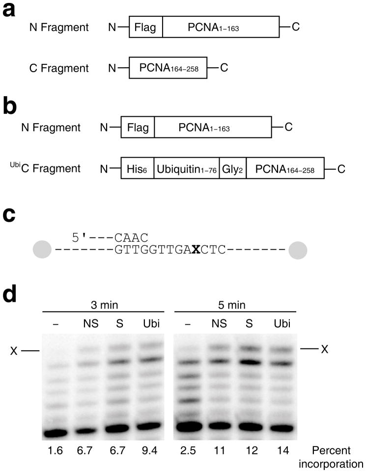
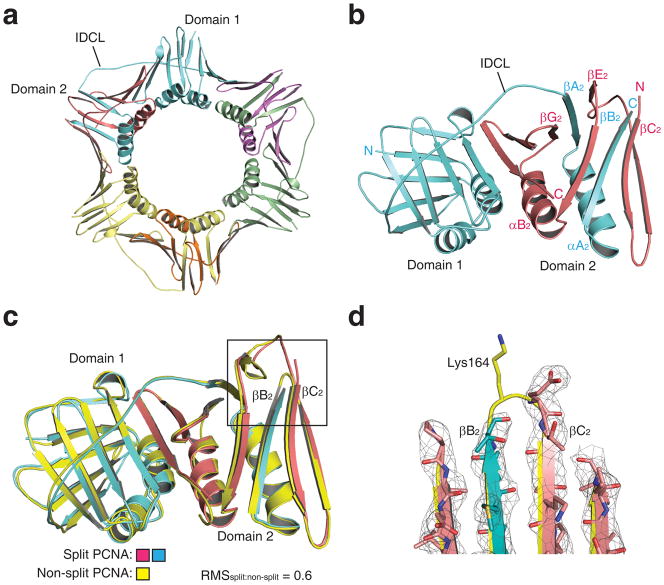
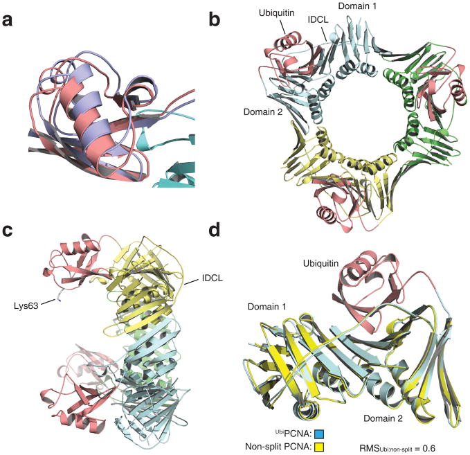
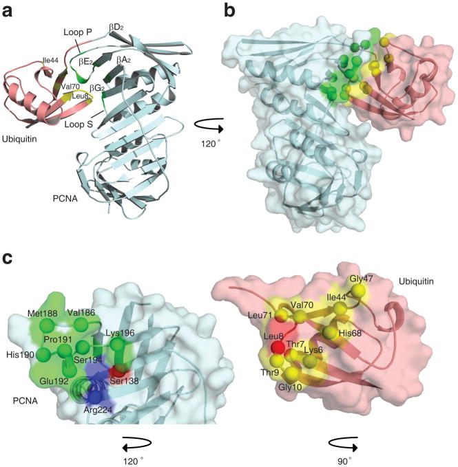
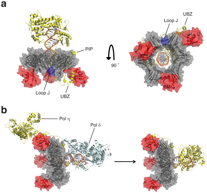
DNA synthesis by classical polymerases can be blocked by many lesions. These blocks are overcome by translesion synthesis, whereby the stalled classical, replicative polymerase is replaced by a nonclassical polymerase. In eukaryotes this polymerase exchange requires proliferating cell nuclear antigen (PCNA) monoubiquitination. To better understand the polymerase exchange, we developed a means of producing monoubiquitinated PCNA, by splitting the protein into two self-assembling polypeptides. We determined the X-ray crystal structure of monoubiquitinated PCNA and found that the ubiquitin moieties are located on the back face of PCNA and interact with it through their canonical hydrophobic surface. Moreover, the attachment of ubiquitin does not change PCNA's conformation. We propose that PCNA ubiquitination facilitates nonclassical polymerase recruitment to the back of PCNA by forming a new binding surface for nonclassical polymerases, consistent with a 'tool belt' model of the polymerase exchange.


Figures

Figure 1
Figure 1
Stimulation of pol η activity by split PCNA and UbiPCNA. (a) Diagram of the two polypeptides used to generate split PCNA is shown. (b) Diagram of the two polypeptides used to generate UbiPCNA is shown. (c) Diagram of the running start DNA substrate used. The ‘X’ represents an abasic site. Both ends of the template strand are capped with biotin-streptavidin blocks. (d) Autoradiograph of the products of the running start reaction of pol η and the indicated DNA substrate after 3 min. and 5 min. The arrow represents incorporation opposite the abasic site. Lanes labeled ‘–’ contain no PCNA, lanes labeled ‘NS’ contain non-split PCNA, lanes labeled ‘S’ contain split PCNA, and lanes labeled ‘Ubi’ contain UbiPCNA. The percent incorporation is shown below each lane.
Figure 2
Figure 2
Viability and UV sensitivity of yeast cells expressing split PCNA and UbiPCNA. (a) The growth of cells producing only non-split PCNA, the K164R mutant PCNA protein, split PCNA, or UbiPCNA is graphed as a function of time. (b) UV sensitivity of cells producing only non-split PCNA, the K164R mutant PCNA protein, split PCNA, or UbiPCNA is shown by graphing the percent of surviving cells as a function of the UV dose. Error bars represent standard deviation.
Figure 3
Figure 3
Structure of split PCNA. (a) Structure of the split PCNA trimer is shown with the three N fragments colored blue, green, and yellow and the three C fragments colored red, purple, and orange. Domain 1, domain 2, and the interdomain connector loop (IDCL) are indicated. (b) Structure of a single split PCNA monomer is shown (viewed from the opposite side of the ring relative to panel a) with the N fragment colored blue and the C fragment colored red. The interdigitating β strands of the two fragments in domain 2 are labeled. (c) The backbone of split PCNA, which is colored blue (N fragment) and red (C fragment), is superimposed on the backbone of non-split PCNA, which is colored yellow. (d) Close up of the loop between β strands βB2 and βC2 showing the position of Lys-164 in non-split PCNA and the break in the backbone between the N fragment and C fragment of split PCNA. The electron density (level=2.0) is shown for split PCNA.
Figure 4
Figure 4
Structure of UbiPCNA. (a) Overlay of the two preferred positions of the ubiquitin moiety. Position 1 is shown in red and position 2 is shown in blue. The corresponding atoms in these positions are separated by 2.5 Å. (b) Structure of the UbiPCNA trimer is shown with the three PCNA subunits shown in blue, green, and yellow and the three ubiquitin moieties (in position 1) shown in red. (c) Side view of the UbiPCNA trimer with the side chain of Lys-63 of the ubiquitin (the site of polyubiquitination) indicated. (d) The backbone of UbiPCNA, which is colored blue (PCNA portion) and red (ubiquitin moiety), is superimposed on the backbone of non-split PCNA, which is colored yellow.
Figure 5
Figure 5
Interactions between ubiquitin and PCNA within UbiPCNA. (a) Ribbon representation showing the ubiquitin-PCNA interface. The ubiquitin moiety is shown in red, and the PCNA is shown in blue. Regions of the ubiquitin moiety contacting the PCNA are shown in yellow, and regions of the PCNA contacting the ubiquitin moiety are shown in green. (b) Space filled representation of the ubiquitin-PCNA interface shown from a different angle. (c) Close up of the interfaces on the ubiquitin moiety and PCNA. The ubiquitin moiety and the PCNA have been separated and rotated relative to the orientation in panel B to show the binding surfaces on each. Residues forming hydrophobic contacts are shown in green and yellow for the PCNA and ubiquitin moiety, respectively. Residues forming electrostatic contacts are shown in blue and red.
Figure 6
Figure 6
Model of the complex between UbiPCNA and pol η. (a) Two views of the model of the UbiPCNA-pol η complex. The PCNA portion is colored grey, loop J of PCNA is colored blue, the ubiquitin moieties are colored red, and the pol η is colored yellow. The PCNA-interacting peptide (PIP) and ubiquitin-binding, zinc-binding (UBZ) motifs of pol η and loop J of PCNA are indicated. (b) A possible tool belt model showing the recruitment of pol η to the side and back face of UbiPCNA while pol δ (colored blue) sits in front of UbiPCNA. Eventually, pol δ is displaced from the front of UbiPCNA by the catalytic core of pol η. For simplicity sake, pol δ is shown as dissociating from the complex, although this need not be the case. The structure of pol δ is from .

References

    1. Prakash S, Sung P, Prakash L. DNA-Repair Genes and Proteins of Saccharomyces-Cerevisiae. Annual Review of Genetics. 1993;27:33–70. - PubMed
    1. Jentsch S, McGrath JP, Varshavsky A. The Yeast DNA-Repair Gene Rad6 Encodes a Ubiquitin-Conjugating Enzyme. Nature. 1987;329:131–134. - PubMed
    1. Bailly V, Lamb J, Sung P, Prakash S, Prakash L. Specific Complex-Formation between Yeast Rad6 and Rad18 Proteins - a Potential Mechanism for Targeting Rad6 Ubiquitin-Conjugating Activity to DNA-Damage Sites. Genes & Development. 1994;8:811–820. - PubMed
    1. Bailly V, Lauder S, Prakash S, Prakash L. Yeast DNA repair proteins Rad6 and Rad18 form a heterodimer that has ubiquitin conjugating, DNA binding, and ATP hydrolytic activities. Journal of Biological Chemistry. 1997;272:23360–23365. - PubMed
    1. Hoege C, Pfander B, Moldovan GL, Pyrowolakis G, Jentsch S. RAD6-dependent DNA repair is linked tomodification of PCNA by ubiquitin and SUMO. Nature. 2002;419:135–141. - PubMed

Publication types

MeSH terms

Substances