. 2010 Apr;16(4):470-4.
doi: 10.1038/nm.2124. Epub 2010 Mar 21.

Myeloperoxidase acts as a profibrotic mediator of atrial fibrillation

Affiliations

Myeloperoxidase acts as a profibrotic mediator of atrial fibrillation

Volker Rudolph et al. Nat Med. 2010 Apr.

Abstract

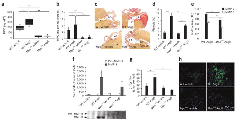
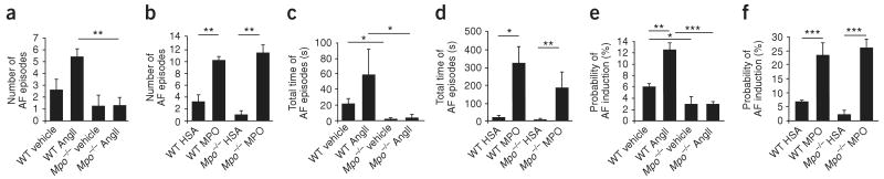
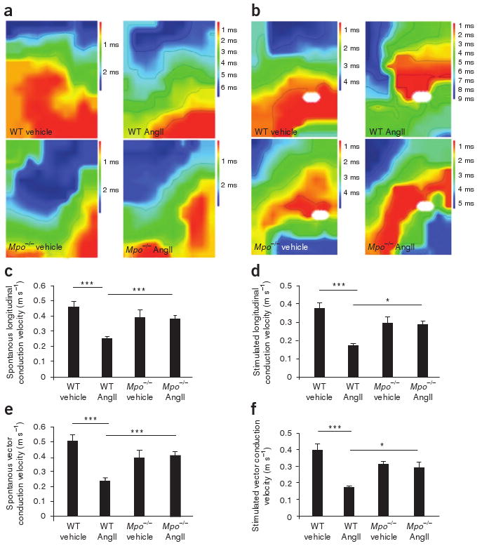
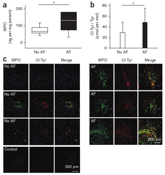
Observational clinical and ex vivo studies have established a strong association between atrial fibrillation and inflammation. However, whether inflammation is the cause or the consequence of atrial fibrillation and which specific inflammatory mediators may increase the atria's susceptibility to fibrillation remain elusive. Here we provide experimental and clinical evidence for the mechanistic involvement of myeloperoxidase (MPO), a heme enzyme abundantly expressed by neutrophils, in the pathophysiology of atrial fibrillation. MPO-deficient mice pretreated with angiotensin II (AngII) to provoke leukocyte activation showed lower atrial tissue abundance of the MPO product 3-chlorotyrosine, reduced activity of matrix metalloproteinases and blunted atrial fibrosis as compared to wild-type mice. Upon right atrial electrophysiological stimulation, MPO-deficient mice were protected from atrial fibrillation, which was reversed when MPO was restored. Humans with atrial fibrillation had higher plasma concentrations of MPO and a larger MPO burden in right atrial tissue as compared to individuals devoid of atrial fibrillation. In the atria, MPO colocalized with markedly increased formation of 3-chlorotyrosine. Our data demonstrate that MPO is a crucial prerequisite for structural remodeling of the myocardium, leading to an increased vulnerability to atrial fibrillation.


Conflict of interest statement

Competing Financial Interests: The authors declare no competing financial interests.

Figures

Figure 1
Figure 1
Mpo−/− mice show less atrial fibrosis, reduced matrix metalloproteinase activity and lower chlorotyrosine bioavailability after AngII treatment. (a,b) MPO plasma concentrations (a) and myocardial MPO deposition (b) in saline (vehicle)- or AngII-treated WT and Mpo−/− mice, as determined by ELISA. n = 14–19, *P < 0.05, **P < 0.01. (c) Quantification of atrial fibrosis in Mpo−/− and WT mice by the picrosirius polarization method. Images are representative of ten images from eight to ten mice per group. LA, left atrium; LV, left ventricle. (d) Quantitative assessment of atrial fibrosis in saline– or AngII-treated WT and Mpo−/− mice. n = 8–12, **P < 0.01, ***P < 0.001. (e) MMP-2 and MMP-9 activity in AngII-treated WT and Mpo−/− mice determined by zymography. n = 7, **P < 0.01. (f) Immunoblot detecting pro–MMP-9 and MMP-9 in mouse atrial tissue. n = 4, P < 0.05 for WT AngII versus Mpo−/− AngII. (g) LC-MS–based quantitative assessment of 3-chlorotyrosine (ClTyr) in atrial tissue of WT and Mpo−/− mice. n = 10, *P < 0.05, ***P < 0.001. (h) Immunostaining of atrial 3-chlorotyrosine in saline– or AngII-treated WT and Mpo−/− mice. All data in b, dg are means ± s.d. Statistical analyses were performed by analysis of variance (ANOVA) followed by Bonferroni's post hoc test. Data in a are presented as median and interquartile range. Kruskal-Wallis test followed by the Mann-Whitney U post hoc test was used for statistical analysis.
Figure 2
Figure 2
Analysis of atrial fibrillation inducibility in Mpo−/− and WT mice in vivo. (ad) After pretreatment with AngII or saline (vehicle) for 14 d (n = 10–16 per group) (a,c) or MPO or human serum albumin (HSA) for 7 d (n = 6–9 per group) (b,d), WT and Mpo−/− mice underwent electrophysiological investigation. (a,b) Quantification of the number of atrial fibrillation (AF) episodes. **P < 0.01. (c,d) Total time of atrial fibrillation episodes. *P < 0.05, **P < 0.01. (e,f) Probability of induction of atrial fibrillation, defined as inducible episodes divided by number of total testing maneuvers applied. *P < 0.05, **P < 0.01, ***P < 0.001. All data are means ± s.d. Statistical analysis was performed by ANOVA followed by Bonferroni's post hoc test.
Figure 3
Figure 3
Epicardial mapping of Langendorff-perfused hearts of AngII- or saline (vehicle)-treated WT or Mpo−/− mice. (a,b) Representative examples of spontaneous (a) and stimulated (b) conduction properties of epicardial activation mapping. White ovals indicate stimulation sites. (cf) Quantification of spontaneous longitudinal conduction velocity (c), stimulated longitudinal conduction velocity (d), spontaneous vector conduction velocity (e) and stimulated vector conduction velocity (f). n = 5–13 per group, *P < 0.05, ***P < 0.001. All data are means ± s.d. Statistical analysis was performed by ANOVA with Bonferroni's post hoc test.
Figure 4
Figure 4
Atrial MPO amounts and protein-bound 3-chlorotyrosine in individuals with or without atrial fibrillation. (a) Quantification of MPO content in tissue homogenates of right atrial appendages of individuals undergoing on-pump coronary bypass surgery, as determined by ELISA normalized to protein concentration (no atrial fibrillation: n = 17, atrial fibrillation: n = 10). Data are presented as median (line) and interquartile range (box); whiskers indicate 5% and 95% percentiles, *P < 0.05. Statistical analyses were performed by the Mann-Whitney U test. (b) LC-MS–based quantitative assessment of 3-chlorotyrosine in atrial tissue of the same individuals. Data are means ± s.d. *P < 0.05. Unpaired Student's t test was used for statistical analysis. (c) Immunofluorescent staining of human right atrial appendage tissue. Left, three representative examples of MPO and the MPO-specific oxidant 3-chlorotyrosine from seven individuals without documented atrial fibrillation. Right, three representative images of right atrial appendage tissue from seven individuals with atrial fibrillation.

References

    1. Issac TT, Dokainish H, Lakkis NM. Role of inflammation in initiation and perpetuation of atrial fibrillation: a systematic review of the published data. J Am Coll Cardiol. 2007;50:2021–2028. - PubMed
    1. Kannel WB, Wolf PA, Benjamin EJ, Levy D. Prevalence, incidence, prognosis and predisposing conditions for atrial fibrillation: population-based estimates. Am J Cardiol. 1998;82:2N–9N. - PubMed
    1. Nattel S, Opie LH. Controversies in atrial fibrillation. Lancet. 2006;367:262–272. - PubMed
    1. Kannel WB, Abbott RD, Savage DD, McNamara PM. Epidemiologic features of chronic atrial fibrillation: the Framingham study. N Engl J Med. 1982;306:1018–1022. - PubMed
    1. Aviles RJ, et al. Inflammation as a risk factor for atrial fibrillation. Circulation. 2003;108:3006–3010. - PubMed

Publication types

MeSH terms